


 癌症的基本特征包括细胞增殖、血管生成、迁移、凋亡逃避机制和细胞永生等。找到癌症发生过程中这些通路的关键标记物和对应的抗体用于检测至关重要。
 为您推荐一个泛素化位点预测神器——泛素化分析工具,可以为您的蛋白的泛素化位点作出预测和评分。
 细胞自噬受体图形绘图工具为你的蛋白的细胞受体结合位点作出预测和评分,识别结合到自噬通路中的蛋白是非常重要的,便于让我们理解自噬在正常生理、病理过程中的作用,如发育、细胞分化、神经退化性疾病、压力条件下、感染和癌症。
p57Kip2 (Mitotic Inhibitor/Suppressor Protein) Antibody - With BSA and Azide
Mouse Monoclonal Antibody [Clone SPM308 ]
- 产品详情
- 实验流程
- 背景知识
Application
| IHC, IF, FC |
|---|---|
| Primary Accession | P49918 |
| Other Accession | 1028, 106070 |
| Reactivity | Human, Mouse |
| Host | Mouse |
| Clonality | Monoclonal |
| Isotype | Mouse / IgG2b, kappa |
| Clone Names | SPM308 |
| Calculated MW | 32177 Da |
| Gene ID | 1028 |
|---|---|
| Other Names | Cyclin-dependent kinase inhibitor 1C, Cyclin-dependent kinase inhibitor p57, p57Kip2, CDKN1C, KIP2 |
| Application Note | IHC~~1:100~500 IF~~1:50~200 FC~~1:10~50 |
| Storage | Store at 2 to 8°C.Antibody is stable for 24 months. |
| Precautions | p57Kip2 (Mitotic Inhibitor/Suppressor Protein) Antibody - With BSA and Azide is for research use only and not for use in diagnostic or therapeutic procedures. |
| Name | CDKN1C |
|---|---|
| Synonyms | KIP2 |
| Function | Potent tight-binding inhibitor of several G1 cyclin/CDK complexes (cyclin E-CDK2, cyclin D2-CDK4, and cyclin A-CDK2) and, to lesser extent, of the mitotic cyclin B-CDC2. Negative regulator of cell proliferation. May play a role in maintenance of the non-proliferative state throughout life. |
| Cellular Location | Nucleus. |
| Tissue Location | Expressed in the heart, brain, lung, skeletal muscle, kidney, pancreas and testis. Expressed in the eye. High levels are seen in the placenta while low levels are seen in the liver |
For Research Use Only. Not For Use In Diagnostic Procedures.
Provided below are standard protocols that you may find useful for product applications.
BACKGROUND
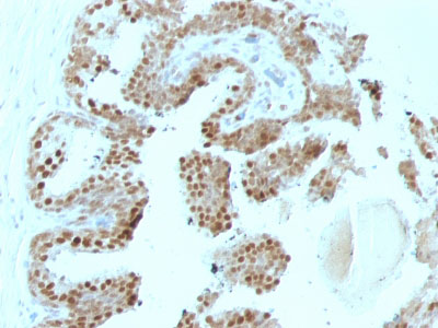
Recognizes a protein of 57kDa, identified as p57Kip2. It shows no cross-reaction with p27Kip1. p57Kip2 is a potent tight-binding inhibitor of several G1 cyclin complexes, and is a negative regulator of cell proliferation. Anti-p57 has been used as an aide in identification of complete hydatidiform mole (CHM) (no nuclear labeling of cytotrophoblasts and stromal cells) from partial hydatidiform mole (PHM) in which both cytotrophoblasts and stromal cells stain. The histological differentiation of complete mole, partial mole, and hydropic spontaneous abortion is problematic. Most complete hydatidiform moles are diploid, whereas most partial moles are triploid. Ploidy studies will identify partial moles, but will not differentiate complete moles from non-molar gestations. Complete moles carry a high risk of persistent disease and choriocarcinoma, while partial moles have a very low risk. In normal placenta, many cytotrophoblast nuclei and stromal cells are labeled with this antibody. Similar findings apply to PHM and hydropic abortus tissues. Intervillous trophoblastic islands (IVTIs) demonstrate nuclear labeling in all three entities and serve as an internal control.
REFERENCES
Lee, M.-H., et al. 1995. Cloning of p57, a cyclin-dependent kinase inhibitor with unique domain structure and tissue distribution. Genes Dev. 9: 639-649. |
终于等到您。ABCEPTA(百远生物)抗体产品。
点击下方“我要评价 ”按钮提交您的反馈信息,您的反馈和评价是我们最宝贵的财富之一,
我们将在1-3个工作日内处理您的反馈信息。
如有疑问,联系:0512-88856768 tech-china@abcepta.com.


