


 癌症的基本特征包括细胞增殖、血管生成、迁移、凋亡逃避机制和细胞永生等。找到癌症发生过程中这些通路的关键标记物和对应的抗体用于检测至关重要。
 为您推荐一个泛素化位点预测神器——泛素化分析工具,可以为您的蛋白的泛素化位点作出预测和评分。
 细胞自噬受体图形绘图工具为你的蛋白的细胞受体结合位点作出预测和评分,识别结合到自噬通路中的蛋白是非常重要的,便于让我们理解自噬在正常生理、病理过程中的作用,如发育、细胞分化、神经退化性疾病、压力条件下、感染和癌症。
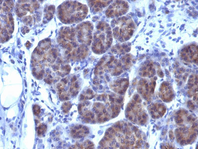
GLG1 (Golgi Glycoprotein 1) (Marker for Human Cells) Antibody - With BSA and Azide
Mouse Monoclonal Antibody [Clone GLG1/970 ]
- 产品详情
- 实验流程
- 背景知识
Application
| WB, IHC, IF, FC, ICC |
|---|---|
| Primary Accession | Q92896 |
| Other Accession | 2734, 109731 |
| Reactivity | Human |
| Host | Mouse |
| Clonality | Monoclonal |
| Isotype | Mouse / IgG1, kappa |
| Clone Names | GLG1/970 |
| Calculated MW | 134552 Da |
| Gene ID | 2734 |
|---|---|
| Other Names | Golgi apparatus protein 1, CFR-1, Cysteine-rich fibroblast growth factor receptor, E-selectin ligand 1, ESL-1, Golgi sialoglycoprotein MG-160, GLG1, CFR1, ESL1, MG160 |
| Application Note | WB~~1:1000 IHC~~1:100~500 IF~~1:50~200 FC~~1:10~50 ICC~~N/A |
| Storage | Store at 2 to 8°C.Antibody is stable for 24 months. |
| Precautions | GLG1 (Golgi Glycoprotein 1) (Marker for Human Cells) Antibody - With BSA and Azide is for research use only and not for use in diagnostic or therapeutic procedures. |
| Name | GLG1 |
|---|---|
| Synonyms | CFR1, ESL1, MG160 |
| Function | Binds fibroblast growth factor and E-selectin (cell-adhesion lectin on endothelial cells mediating the binding of neutrophils). |
| Cellular Location | Golgi apparatus membrane; Single-pass type I membrane protein. Golgi outpost {ECO:0000250|UniProtKB:Q62638}. Cytoplasm, cytoskeleton, microtubule organizing center {ECO:0000250|UniProtKB:Q62638}. Note=Golgi medial cisternae. Localizes to the postsynaptic Golgi apparatus region, also named Golgi outpost, which shapes dendrite morphology by functioning as sites of acentrosomal microtubule nucleation. {ECO:0000250|UniProtKB:Q62638} |
| Tissue Location | Widely expressed. Highest levels in pancreas, skeletal muscle, placenta, heart, testis and ovary. Also found in the kidney, liver, lung and brain. |
For Research Use Only. Not For Use In Diagnostic Procedures.
Provided below are standard protocols that you may find useful for product applications.
BACKGROUND
This MAb recognizes a protein of 134kDa, which binds fibroblast growth factor and E-selectin (cell-adhesion lectin on endothelial cells mediating the binding of neutrophils). Fucosylation is essential for binding to E-selectin. It contains sialic acid residues and 16 Cys-rich GLG1 repeats. This MAb can be used to stain the Golgi complex in cell or tissue preparations and can be used as a Golgi marker in subcellular fractions. It produces a diffuse staining pattern of the Golgi zone in normal and malignant cells. This MAb is an excellent marker for human cells in xenographic model research. It reacts specifically with human cells. The Golgi apparatus is an organelle present in all eukaryotic cells that forms a part of the endomembrane system. The primary function of the Golgi apparatus is to process and package macromolecules synthesized by the cell for exocytosis or use within the cell. The Golgi is made up of a stack of flattened, membrane-bound sacs known as cisternae, with three functional regions: the cis face, medial region and trans face. Each region consists of various enzymes that selectively modify the macromolecules passing though them, depending on where they are destined to reside. Several spherical vesicles that have budded off of the Golgi are present surrounding the main cisternae. The Golgi tends to be more pronounced and numerous in cells that make and secrete many substances such as plasma B cells.
REFERENCES
Nakamura et. al. 1995. J. Cell Biol. 131:1715-26. | Nakamura et.al. 1997. Cell. 89(3):445-55
终于等到您。ABCEPTA(百远生物)抗体产品。
点击下方“我要评价 ”按钮提交您的反馈信息,您的反馈和评价是我们最宝贵的财富之一,
我们将在1-3个工作日内处理您的反馈信息。
如有疑问,联系:0512-88856768 tech-china@abcepta.com.


