


 癌症的基本特征包括细胞增殖、血管生成、迁移、凋亡逃避机制和细胞永生等。找到癌症发生过程中这些通路的关键标记物和对应的抗体用于检测至关重要。
 为您推荐一个泛素化位点预测神器——泛素化分析工具,可以为您的蛋白的泛素化位点作出预测和评分。
 细胞自噬受体图形绘图工具为你的蛋白的细胞受体结合位点作出预测和评分,识别结合到自噬通路中的蛋白是非常重要的,便于让我们理解自噬在正常生理、病理过程中的作用,如发育、细胞分化、神经退化性疾病、压力条件下、感染和癌症。
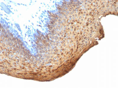
Cytokeratin 10/13 Antibody - With BSA and Azide
Mouse Monoclonal Antibody [Clone DE-K13 ]
- 产品详情
- 实验流程
- 背景知识
Application
| WB, IF, FC, IHC-P |
|---|---|
| Primary Accession | P13645 |
| Other Accession | 3858, 99936 |
| Reactivity | Human, Cat |
| Host | Mouse |
| Clonality | Monoclonal |
| Isotype | Mouse / IgG2a, kappa |
| Clone Names | DE-K13 |
| Calculated MW | 58827 Da |
| Gene ID | 3858 |
|---|---|
| Other Names | Keratin, type I cytoskeletal 10, Cytokeratin-10, CK-10, Keratin-10, K10, KRT10, KPP |
| Application Note | WB~~1:1000 IF~~1:50~200 FC~~1:10~50 IHC-P~~N/A |
| Storage | Store at 2 to 8°C.Antibody is stable for 24 months. |
| Precautions | Cytokeratin 10/13 Antibody - With BSA and Azide is for research use only and not for use in diagnostic or therapeutic procedures. |
| Name | KRT10 |
|---|---|
| Synonyms | KPP |
| Function | Plays a role in the establishment of the epidermal barrier on plantar skin (By similarity). Involved in the maintenance of cell layer development and keratin filament bundles in suprabasal cells of the epithelium (By similarity). |
| Cellular Location | Secreted, extracellular space. Cell surface. Cytoplasm |
| Tissue Location | Seen in all suprabasal cell layers including stratum corneum. Expressed on the surface of lung cell lines (PubMed:19627498). Localized on the surface of desquamated nasal epithelial cells (at protein level) (PubMed:12427098) |
For Research Use Only. Not For Use In Diagnostic Procedures.
Provided below are standard protocols that you may find useful for product applications.
BACKGROUND
This antibody recognizes cytokeratin 10 (56.5kDa) and cytokeratin 13 (53kDa) in Western blotting. It recognizes only cytokeratin 13 in formalin-fixed, paraffin-embedded tissue sections. It does not react with cytokeratin 10 positive, cytokeratin 13 negative epithelia such as epidermis. However, on frozen sections this MAb serves as differentiation-related marker of all stratified epithelia; it stains all suprabasal cells in both cornifying and non-cornifying stratified epithelia and more differentiated cells of squamous carcinomas.
REFERENCES
Ivanyi D et. al. Journal of Pathology, 1989, 159:7-12.,Ivanyi, D., Minke, J. M., Hageman, C., Groeneveld, E., and van Doornewaard, G. (1992). Patterns of expression of feline cytokeratins in healthy epithelia and mammary carcinoma cells, Am J Vet Res 53, 304-14. ,Ivanyi, D., Minke, J. M., Hageman, C., Groeneveld, E., van Doornewaard, G., and Misdorp, W. (1993). Cytokeratins as markers of initial stages of squamous metaplasia in feline mammary carcinomas, Am J Vet Res 54, 1095
终于等到您。ABCEPTA(百远生物)抗体产品。
点击下方“我要评价 ”按钮提交您的反馈信息,您的反馈和评价是我们最宝贵的财富之一,
我们将在1-3个工作日内处理您的反馈信息。
如有疑问,联系:0512-88856768 tech-china@abcepta.com.


