


 癌症的基本特征包括细胞增殖、血管生成、迁移、凋亡逃避机制和细胞永生等。找到癌症发生过程中这些通路的关键标记物和对应的抗体用于检测至关重要。
 为您推荐一个泛素化位点预测神器——泛素化分析工具,可以为您的蛋白的泛素化位点作出预测和评分。
 细胞自噬受体图形绘图工具为你的蛋白的细胞受体结合位点作出预测和评分,识别结合到自噬通路中的蛋白是非常重要的,便于让我们理解自噬在正常生理、病理过程中的作用,如发育、细胞分化、神经退化性疾病、压力条件下、感染和癌症。
RET Antibody (Ascites)
Mouse Monoclonal Antibody (Mab)
- 产品详情
- 文献引用 : 1
- 实验流程
- 背景知识
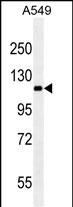
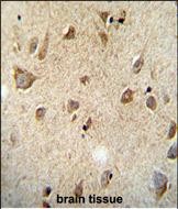
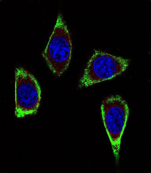
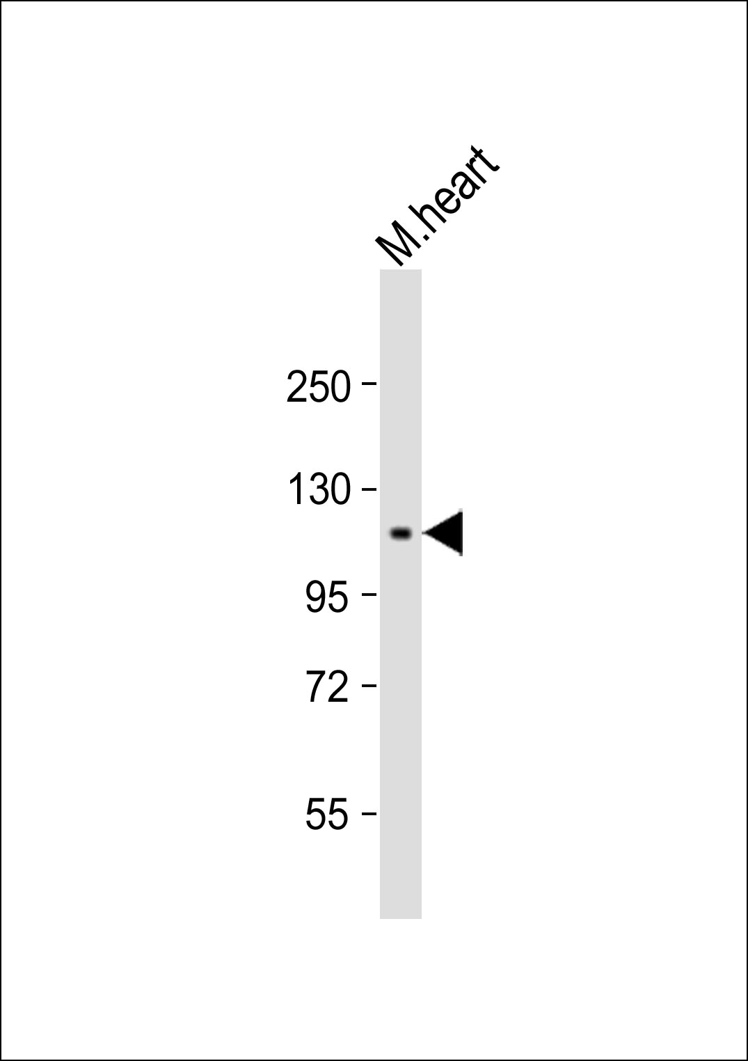
Application
| WB, IHC-P, IF, E |
|---|---|
| Primary Accession | P07949 |
| Other Accession | NP_065681.1, NP_066124.1 |
| Reactivity | Human |
| Host | Mouse |
| Clonality | Monoclonal |
| Isotype | IgM,K |
| Clone Names | 188CT11.2.3 |
| Calculated MW | 124319 Da |
| Gene ID | 5979 |
|---|---|
| Other Names | Proto-oncogene tyrosine-protein kinase receptor Ret, Cadherin family member 12, Proto-oncogene c-Ret, Soluble RET kinase fragment, Extracellular cell-membrane anchored RET cadherin 120 kDa fragment, RET, CDHF12, CDHR16, PTC, RET51 |
| Target/Specificity | This RET Monoclonal antibody was raised using purified His-tagged recombinant human RET. |
| Dilution | WB~~ 1:1000 IHC-P~~1:100~500 IF~~1:10~50 E~~Use at an assay dependent concentration. |
| Format | Mouse monoclonal antibody supplied in crude ascites with 0.09% (W/V) sodium azide. |
| Storage | Maintain refrigerated at 2-8°C for up to 2 weeks. For long term storage store at -20°C in small aliquots to prevent freeze-thaw cycles. |
| Precautions | RET Antibody (Ascites) is for research use only and not for use in diagnostic or therapeutic procedures. |
| Name | RET {ECO:0000303|PubMed:2660074, ECO:0000312|HGNC:HGNC:9967} |
|---|---|
| Function | Receptor tyrosine-protein kinase involved in numerous cellular mechanisms including cell proliferation, neuronal navigation, cell migration, and cell differentiation in response to glia cell line- derived growth family factors (GDNF, NRTN, ARTN, PSPN and GDF15) (PubMed:20064382, PubMed:20616503, PubMed:20702524, PubMed:21357690, PubMed:21454698, PubMed:24560924, PubMed:28846097, PubMed:28846099, PubMed:28953886, PubMed:31118272). In contrast to most receptor tyrosine kinases, RET requires not only its cognate ligands but also coreceptors, for activation (PubMed:21994944, PubMed:23333276, PubMed:28846097, PubMed:28846099, PubMed:28953886). GDNF ligands (GDNF, NRTN, ARTN, PSPN and GDF15) first bind their corresponding GDNFR coreceptors (GFRA1, GFRA2, GFRA3, GFRA4 and GFRAL, respectively), triggering RET autophosphorylation and activation, leading to activation of downstream signaling pathways, including the MAPK- and AKT-signaling pathways (PubMed:21994944, PubMed:23333276, PubMed:24560924, PubMed:25242331, PubMed:28846097, PubMed:28846099, PubMed:28953886). Acts as a dependence receptor via the GDNF-GFRA1 signaling: in the presence of the ligand GDNF in somatotrophs within pituitary, promotes survival and down regulates growth hormone (GH) production, but triggers apoptosis in absence of GDNF (PubMed:20616503, PubMed:21994944). Required for the molecular mechanisms orchestration during intestine organogenesis via the ARTN-GFRA3 signaling: involved in the development of enteric nervous system and renal organogenesis during embryonic life, and promotes the formation of Peyer's patch-like structures, a major component of the gut-associated lymphoid tissue (By similarity). Mediates, through interaction with GDF15-receptor GFRAL, GDF15-induced cell-signaling in the brainstem which triggers an aversive response, characterized by nausea, vomiting, and/or loss of appetite in response to various stresses (PubMed:28846097, PubMed:28846099, PubMed:28953886). Modulates cell adhesion via its cleavage by caspase in sympathetic neurons and mediates cell migration in an integrin (e.g. ITGB1 and ITGB3)-dependent manner (PubMed:20702524, PubMed:21357690). Also active in the absence of ligand, triggering apoptosis through a mechanism that requires receptor intracellular caspase cleavage (PubMed:21357690). Triggers the differentiation of rapidly adapting (RA) mechanoreceptors (PubMed:20064382). Involved in the development of the neural crest (By similarity). Regulates nociceptor survival and size (By similarity). Phosphorylates PTK2/FAK1 (PubMed:21454698). |
| Cellular Location | Cell membrane; Single-pass type I membrane protein. Endosome membrane; Single-pass type I membrane protein Note=Predominantly located on the plasma membrane (PubMed:23333276, PubMed:9575150). In the presence of SORL1 and GFRA1, directed to endosomes (PubMed:23333276). |
For Research Use Only. Not For Use In Diagnostic Procedures.

Provided below are standard protocols that you may find useful for product applications.
BACKGROUND
This gene, a member of the cadherin superfamily, encodes one of the receptor tyrosine kinases, which are cell-surface molecules that transduce signals for cell growth and differentiation. This gene plays a crucial role in neural crest development, and it can undergo oncogenic activation in vivo and in vitro by cytogenetic rearrangement. Mutations in this gene are associated with the disorders multiple endocrine neoplasia, type IIA, multiple endocrine neoplasia, type IIB, Hirschsprung disease, and medullary thyroid carcinoma. Two transcript variants encoding different isoforms have been found for this gene. Additional transcript variants have been described but their biological validity has not been confirmed.
REFERENCES
Siqueira, D.R., et al. Endocr. Relat. Cancer 17(4):953-963(2010)
Gockel, H.R., et al. Hum. Genet. 128(4):353-364(2010)
Kim, H.K., et al. Anticancer Res. 30(9):3621-3627(2010)
Pacini, F., et al. Clin Oncol (R Coll Radiol) 22(6):475-485(2010)
Jugessur, A., et al. PLoS ONE 5 (7), E11493 (2010) :
终于等到您。ABCEPTA(百远生物)抗体产品。
点击下方“我要评价 ”按钮提交您的反馈信息,您的反馈和评价是我们最宝贵的财富之一,
我们将在1-3个工作日内处理您的反馈信息。
如有疑问,联系:0512-88856768 tech-china@abcepta.com.


