


 癌症的基本特征包括细胞增殖、血管生成、迁移、凋亡逃避机制和细胞永生等。找到癌症发生过程中这些通路的关键标记物和对应的抗体用于检测至关重要。
 为您推荐一个泛素化位点预测神器——泛素化分析工具,可以为您的蛋白的泛素化位点作出预测和评分。
 细胞自噬受体图形绘图工具为你的蛋白的细胞受体结合位点作出预测和评分,识别结合到自噬通路中的蛋白是非常重要的,便于让我们理解自噬在正常生理、病理过程中的作用,如发育、细胞分化、神经退化性疾病、压力条件下、感染和癌症。
RAB3A Antibody
Purified Mouse Monoclonal Antibody (Mab)
- 产品详情
- 文献引用 : 1
- 实验流程
- 背景知识
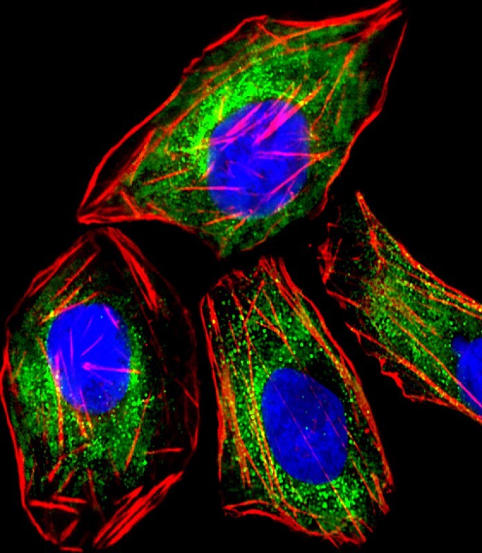
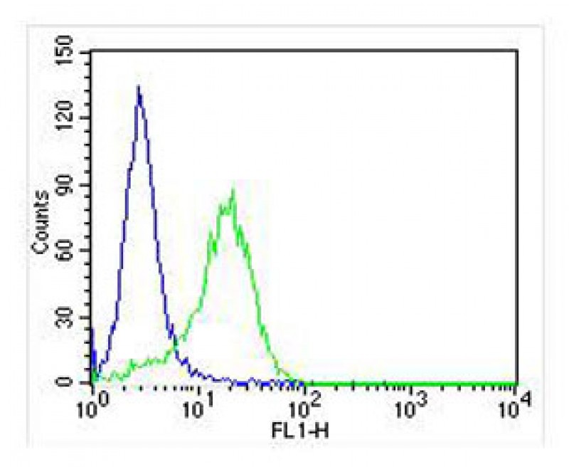
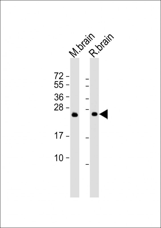
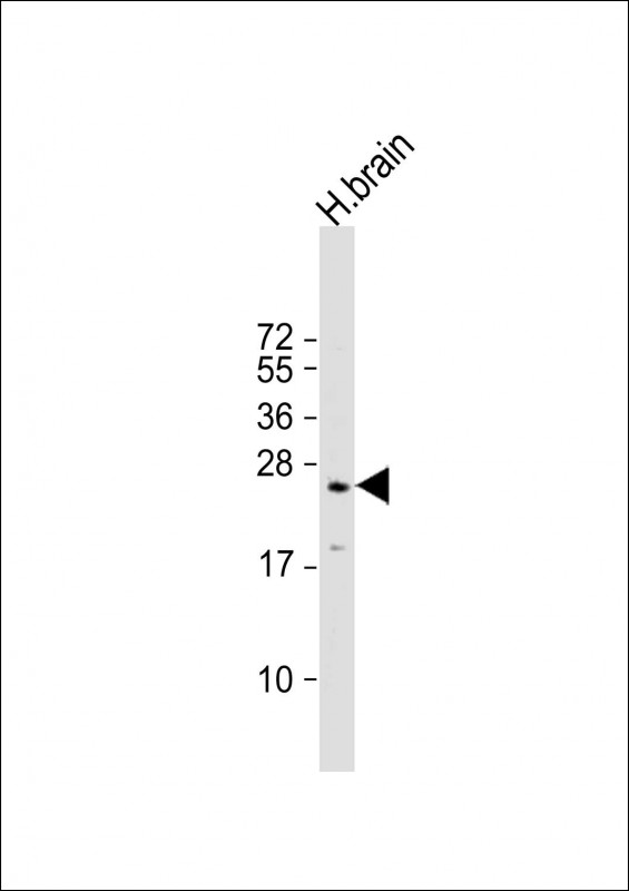
Application
| WB, FC, IF, E |
|---|---|
| Primary Accession | P20336 |
| Reactivity | Human, Rat, Mouse |
| Host | Mouse |
| Clonality | monoclonal |
| Isotype | IgG1,k |
| Clone Names | 1531CT562.14.57 |
| Calculated MW | 24984 Da |
| Gene ID | 5864 |
|---|---|
| Other Names | Ras-related protein Rab-3A, RAB3A |
| Target/Specificity | This RAB3A antibody is generated from a mouse immunized with a recombinant protein. |
| Dilution | WB~~1:500 FC~~1:25 IF~~1:25 E~~Use at an assay dependent concentration. |
| Format | Purified monoclonal antibody supplied in PBS with 0.09% (W/V) sodium azide. This antibody is purified through a protein G column, followed by dialysis against PBS. |
| Storage | Maintain refrigerated at 2-8°C for up to 2 weeks. For long term storage store at -20°C in small aliquots to prevent freeze-thaw cycles. |
| Precautions | RAB3A Antibody is for research use only and not for use in diagnostic or therapeutic procedures. |
| Name | RAB3A (HGNC:9777) |
|---|---|
| Function | The small GTPases Rab are key regulators of intracellular membrane trafficking, from the formation of transport vesicles to their fusion with membranes (PubMed:2501306). Rabs cycle between an inactive GDP-bound form and an active GTP-bound form that is able to recruit to membranes different sets of downstream effectors directly responsible for vesicle formation, movement, tethering and fusion (PubMed:2501306). RAB3A plays a central role in regulated exocytosis and secretion. Controls the recruitment, tethering and docking of secretory vesicles to the plasma membrane (PubMed:2501306). Upon stimulation, switches to its active GTP-bound form, cycles to vesicles and recruits effectors such as RIMS1, RIMS2, Rabphilin-3A/RPH3A, RPH3AL or SYTL4 to help the docking of vesicules onto the plasma membrane (By similarity). Upon GTP hydrolysis by GTPase-activating protein, dissociates from the vesicle membrane allowing the exocytosis to proceed (By similarity). Stimulates insulin secretion through interaction with RIMS2 or RPH3AL effectors in pancreatic beta cells (By similarity). Regulates calcium-dependent lysosome exocytosis and plasma membrane repair (PMR) via the interaction with 2 effectors, SYTL4 and myosin-9/MYH9 (PubMed:27325790). Acts as a positive regulator of acrosome content secretion in sperm cells by interacting with RIMS1 (PubMed:22248876, PubMed:30599141). Also plays a role in the regulation of dopamine release by interacting with synaptotagmin I/SYT (By similarity). |
| Cellular Location | Cytoplasm, cytosol {ECO:0000250|UniProtKB:P63012}. Lysosome Cytoplasmic vesicle, secretory vesicle {ECO:0000250|UniProtKB:P63012} Cell projection, axon {ECO:0000250|UniProtKB:P63011}. Cell membrane; Lipid-anchor; Cytoplasmic side. Presynapse {ECO:0000250|UniProtKB:P63011}. Postsynapse {ECO:0000250|UniProtKB:P63011}. Note=Cycles between a vesicle- associated GTP-bound form and a cytosolic GDP-bound form {ECO:0000250|UniProtKB:P63012} |
| Tissue Location | Specifically expressed in brain. |
For Research Use Only. Not For Use In Diagnostic Procedures.

Provided below are standard protocols that you may find useful for product applications.
BACKGROUND
Involved in exocytosis by regulating a late step in synaptic vesicle fusion. Could play a role in neurotransmitter release by regulating membrane flow in the nerve terminal.
REFERENCES
Zahraoui A.,et al.J. Biol. Chem. 264:12394-12401(1989).
Sullivan M.,et al.Cell. Signal. 11:735-742(1999).
Liu Y.,et al.Submitted (APR-2000) to the EMBL/GenBank/DDBJ databases.
Puhl H.L. III,et al.Submitted (APR-2002) to the EMBL/GenBank/DDBJ databases.
Ota T.,et al.Nat. Genet. 36:40-45(2004).
终于等到您。ABCEPTA(百远生物)抗体产品。
点击下方“我要评价 ”按钮提交您的反馈信息,您的反馈和评价是我们最宝贵的财富之一,
我们将在1-3个工作日内处理您的反馈信息。
如有疑问,联系:0512-88856768 tech-china@abcepta.com.


