


 癌症的基本特征包括细胞增殖、血管生成、迁移、凋亡逃避机制和细胞永生等。找到癌症发生过程中这些通路的关键标记物和对应的抗体用于检测至关重要。
 为您推荐一个泛素化位点预测神器——泛素化分析工具,可以为您的蛋白的泛素化位点作出预测和评分。
 细胞自噬受体图形绘图工具为你的蛋白的细胞受体结合位点作出预测和评分,识别结合到自噬通路中的蛋白是非常重要的,便于让我们理解自噬在正常生理、病理过程中的作用,如发育、细胞分化、神经退化性疾病、压力条件下、感染和癌症。
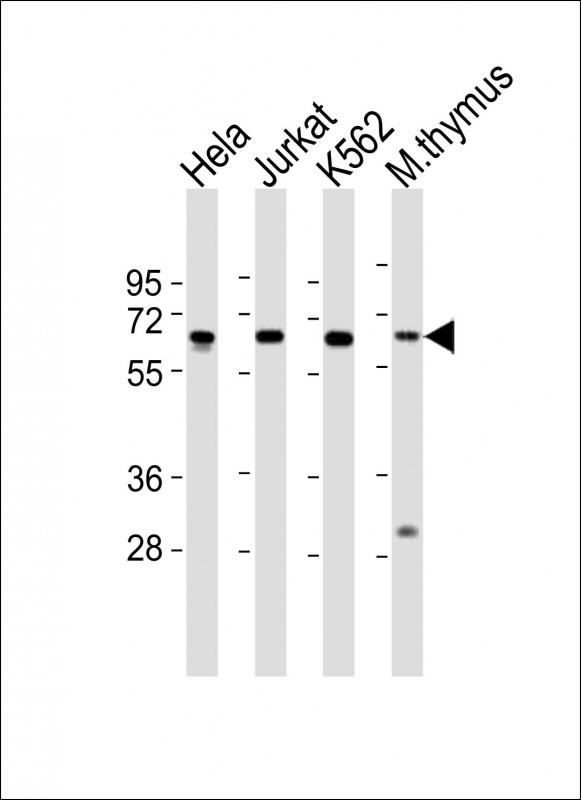
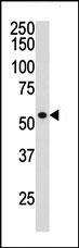
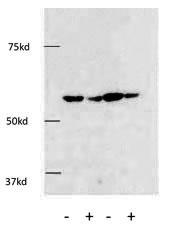
HDAC1 Antibody (C-term)
Purified Rabbit Polyclonal Antibody (Pab)
- 产品详情
- 文献引用 : 1
- 实验流程
- 背景知识
Application
| WB, IF, E |
|---|---|
| Primary Accession | Q13547 |
| Other Accession | O09106 |
| Reactivity | Human, Rat, Mouse |
| Predicted | Mouse |
| Host | Rabbit |
| Clonality | Polyclonal |
| Isotype | Rabbit IgG |
| Calculated MW | 55103 Da |
| Antigen Region | 449-482 aa |
| Gene ID | 3065 |
|---|---|
| Other Names | Histone deacetylase 1, HD1, HDAC1, RPD3L1 |
| Target/Specificity | This HDAC1 antibody is generated from rabbits immunized with a KLH conjugated synthetic peptide between 449-482 amino acids from the C-terminal region of human HDAC1. |
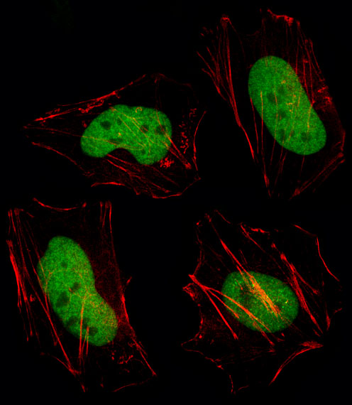
| Dilution | WB~~1:1000 IF~~1:10~50 E~~Use at an assay dependent concentration. |
| Format | Purified polyclonal antibody supplied in PBS with 0.09% (W/V) sodium azide. This antibody is prepared by Saturated Ammonium Sulfate (SAS) precipitation followed by dialysis against PBS. |
| Storage | Maintain refrigerated at 2-8°C for up to 2 weeks. For long term storage store at -20°C in small aliquots to prevent freeze-thaw cycles. |
| Precautions | HDAC1 Antibody (C-term) is for research use only and not for use in diagnostic or therapeutic procedures. |
| Name | HDAC1 {ECO:0000303|PubMed:10846170, ECO:0000312|HGNC:HGNC:4852} |
|---|---|
| Function | Histone deacetylase that catalyzes the deacetylation of lysine residues on the N-terminal part of the core histones (H2A, H2B, H3 and H4) (PubMed:16762839, PubMed:17704056, PubMed:28497810). Histone deacetylation gives a tag for epigenetic repression and plays an important role in transcriptional regulation, cell cycle progression and developmental events (PubMed:16762839, PubMed:17704056). Histone deacetylases act via the formation of large multiprotein complexes (PubMed:16762839, PubMed:17704056). Acts as a component of the histone deacetylase NuRD complex which participates in the remodeling of chromatin (PubMed:16428440, PubMed:28977666). As part of the SIN3B complex is recruited downstream of the constitutively active genes transcriptional start sites through interaction with histones and mitigates histone acetylation and RNA polymerase II progression within transcribed regions contributing to the regulation of transcription (PubMed:21041482). Also functions as a deacetylase for non-histone targets, such as NR1D2, RELA, SP1, SP3, STAT3, ZNF76 and TSHZ3 (PubMed:12837748, PubMed:16285960, PubMed:16337145, PubMed:16478997, PubMed:17996965, PubMed:19343227). Deacetylates SP proteins, SP1 and SP3, and regulates their function (PubMed:12837748, PubMed:16478997). Component of the BRG1-RB1-HDAC1 complex, which negatively regulates the CREST-mediated transcription in resting neurons (PubMed:19081374). Upon calcium stimulation, HDAC1 is released from the complex and CREBBP is recruited, which facilitates transcriptional activation (PubMed:19081374). Deacetylates TSHZ3 and regulates its transcriptional repressor activity (PubMed:19343227). Deacetylates 'Lys-310' in RELA and thereby inhibits the transcriptional activity of NF-kappa-B (PubMed:17000776). Deacetylates NR1D2 and abrogates the effect of KAT5- mediated relieving of NR1D2 transcription repression activity (PubMed:17996965). Component of a RCOR/GFI/KDM1A/HDAC complex that suppresses, via histone deacetylase (HDAC) recruitment, a number of genes implicated in multilineage blood cell development (By similarity). Involved in CIART-mediated transcriptional repression of the circadian transcriptional activator: CLOCK-BMAL1 heterodimer (By similarity). Required for the transcriptional repression of circadian target genes, such as PER1, mediated by the large PER complex or CRY1 through histone deacetylation (By similarity). In addition to protein deacetylase activity, also has protein-lysine deacylase activity: acts as a protein decrotonylase and delactylase by mediating decrotonylation ((2E)-butenoyl) and delactylation (lactoyl) of histones, respectively (PubMed:28497810, PubMed:35044827). |
| Cellular Location | Nucleus |
| Tissue Location | Ubiquitous, with higher levels in heart, pancreas and testis, and lower levels in kidney and brain |
For Research Use Only. Not For Use In Diagnostic Procedures.

Provided below are standard protocols that you may find useful for product applications.
BACKGROUND
Histone acetylation and deacetylation, catalyzed by multisubunit complexes, play a key role in the regulation of eukaryotic gene expression. HDAC1 belongs to the histone deacetylase/acuc/apha family and is a component of the histone deacetylase complex. It also interacts with retinoblastoma tumor-suppressor protein and this complex is a key element in the control of cell proliferation and differentiation. Together with metastasis-associated protein-2, it deacetylates p53 and modulates its effect on cell growth and apoptosis.
REFERENCES
Di Padova, M., et al., J. Biol. Chem. 278(38):36496-36504 (2003).
Wang, S., et al., Oncogene 22(40):6204-6213 (2003).
Xia, Z.B., et al., Proc. Natl. Acad. Sci. U.S.A. 100(14):8342-8347 (2003).
Rocha, S., et al., Mol. Cell. Biol. 23(13):4713-4727 (2003).
Macaluso, M., et al., Oncogene 22(23):3511-3517 (2003).
终于等到您。ABCEPTA(百远生物)抗体产品。
点击下方“我要评价 ”按钮提交您的反馈信息,您的反馈和评价是我们最宝贵的财富之一,
我们将在1-3个工作日内处理您的反馈信息。
如有疑问,联系:0512-88856768 tech-china@abcepta.com.


