


 癌症的基本特征包括细胞增殖、血管生成、迁移、凋亡逃避机制和细胞永生等。找到癌症发生过程中这些通路的关键标记物和对应的抗体用于检测至关重要。
 为您推荐一个泛素化位点预测神器——泛素化分析工具,可以为您的蛋白的泛素化位点作出预测和评分。
 细胞自噬受体图形绘图工具为你的蛋白的细胞受体结合位点作出预测和评分,识别结合到自噬通路中的蛋白是非常重要的,便于让我们理解自噬在正常生理、病理过程中的作用,如发育、细胞分化、神经退化性疾病、压力条件下、感染和癌症。
Mouse TLR2 Antibody (C-term)
Purified Rabbit Polyclonal Antibody (Pab)
- 产品详情
- 文献引用 : 1
- 实验流程
- 背景知识
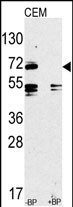
Application
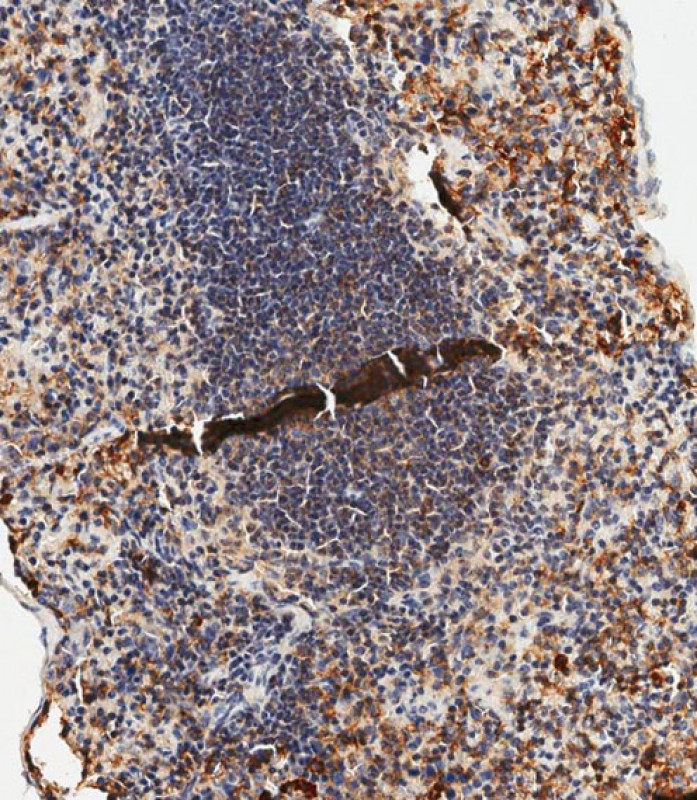
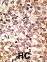
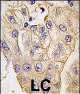
| WB, IHC-P-Leica, E |
|---|---|
| Primary Accession | Q9QUN7 |
| Reactivity | Human, Mouse |
| Host | Rabbit |
| Clonality | Polyclonal |
| Isotype | Rabbit IgG |
| Calculated MW | 89449 Da |
| Antigen Region | 720-749 aa |
| Gene ID | 24088 |
|---|---|
| Other Names | Toll-like receptor 2, CD282, Tlr2 |
| Target/Specificity | This Mouse TLR2 antibody is generated from rabbits immunized with a KLH conjugated synthetic peptide between 720-749 amino acids from the C-terminal region of mouse TLR2. |
| Dilution | WB~~1:1000 IHC-P-Leica~~1:500 E~~Use at an assay dependent concentration. |
| Format | Purified polyclonal antibody supplied in PBS with 0.09% (W/V) sodium azide. This antibody is prepared by Saturated Ammonium Sulfate (SAS) precipitation followed by dialysis against PBS. |
| Storage | Maintain refrigerated at 2-8°C for up to 2 weeks. For long term storage store at -20°C in small aliquots to prevent freeze-thaw cycles. |
| Precautions | Mouse TLR2 Antibody (C-term) is for research use only and not for use in diagnostic or therapeutic procedures. |
| Name | Tlr2 |
|---|---|
| Function | Cooperates with LY96 to mediate the innate immune response to bacterial lipoproteins and other microbial cell wall components. Cooperates with TLR1 or TLR6 to mediate the innate immune response to bacterial lipoproteins or lipopeptides. Acts via MYD88 and TRAF6, leading to NF-kappa-B activation, cytokine secretion and the inflammatory response (By similarity) (PubMed:15690042). May also promote apoptosis in response to lipoproteins (By similarity). Forms activation clusters composed of several receptors depending on the ligand, these clusters trigger signaling from the cell surface and subsequently are targeted to the Golgi in a lipid-raft dependent pathway. Forms the cluster TLR2:TLR6:CD14:CD36 in response to diacylated lipopeptides and TLR2:TLR1:CD14 in response to triacylated lipopeptides (By similarity). Recognizes M.tuberculosis major T-antigen EsxA (ESAT-6) which inhibits downstream MYD88-dependent signaling (PubMed:17486091). Acts as the major receptor for M.tuberculosis lipoproteins LprA, LprG, LpqH and PhoS1 (pstS1), in conjunction with TLR1 and for some but not all lipoproteins CD14 and/or CD36. The lipoproteins act as agonists to modulate antigen presenting cell functions in response to the pathogen (PubMed:19362712). Recombinant MPT83 from M.tuberculosis stimulates secretion of cytokines (TNF, IL-6 and IL-12p40) by mouse macrophage cell lines in a TLR2-dependent fashion, which leads to increased host innate immunity responses against the bacterium (PubMed:22174456). Lung macrophages which express low levels of TLR2 respond poorly to stimulation by M.tuberculosis LpqH (PubMed:19362712). Required for normal uptake of M.tuberculosis, a process that is inhibited by M.tuberculosis LppM (PubMed:27220037). Interacts with TICAM2 (By similarity). |
| Cellular Location | Cell membrane; Single-pass type I membrane protein. Cytoplasmic vesicle, phagosome membrane; Single-pass type I membrane protein. Membrane raft {ECO:0000250|UniProtKB:O60603}. Note=Does not reside in lipid rafts before stimulation but accumulates increasingly in the raft upon the presence of the microbial ligand. In response to diacylated lipoproteins, TLR2:TLR6 heterodimers are recruited in lipid rafts, this recruitment determine the intracellular targeting to the Golgi apparatus. Triacylated lipoproteins induce the same mechanism for TLR2:TLR1 heterodimers. {ECO:0000250|UniProtKB:O60603} |
| Tissue Location | Detected in a macrophage cell line, smooth muscle, lung, spleen, thymus, brain and adipose tissue. Cell surface expression detected in lung alveolar macrophages, dendritic macrophages and at lower levels in lung macrophages (at protein level) (PubMed:19362712) |
For Research Use Only. Not For Use In Diagnostic Procedures.

Provided below are standard protocols that you may find useful for product applications.
BACKGROUND
TLR2 is a member of the Toll-like receptor (TLR) family which plays a fundamental role in pathogen recognition and activation of innate immunity. TLRs are highly conserved from Drosophila to humans and share structural and functional similarities. They recognize pathogen-associated molecular patterns (PAMPs) that are expressed on infectious agents, and mediate the production of cytokines necessary for the development of effective immunity. The various TLRs exhibit different patterns of expression. TLR2 is expressed most abundantly in peripheral blood leukocytes, and mediates host response to Gram-positive bacteria and yeast via stimulation of NF-kappaB.
REFERENCES
Okazaki, Y., et al., Nature 420(6915):563-573 (2002). Lin, Y., et al., J. Biol. Chem. 275(32):24255-24263 (2000). Matsuguchi, T., et al., Blood 95(4):1378-1385 (2000). Heine, H., et al., J. Immunol. 162(12):6971-6975 (1999). Underhill, D.M., et al., Nature 401(6755):811-815 (1999).
终于等到您。ABCEPTA(百远生物)抗体产品。
点击下方“我要评价 ”按钮提交您的反馈信息,您的反馈和评价是我们最宝贵的财富之一,
我们将在1-3个工作日内处理您的反馈信息。
如有疑问,联系:0512-88856768 tech-china@abcepta.com.


