


 癌症的基本特征包括细胞增殖、血管生成、迁移、凋亡逃避机制和细胞永生等。找到癌症发生过程中这些通路的关键标记物和对应的抗体用于检测至关重要。
 为您推荐一个泛素化位点预测神器——泛素化分析工具,可以为您的蛋白的泛素化位点作出预测和评分。
 细胞自噬受体图形绘图工具为你的蛋白的细胞受体结合位点作出预测和评分,识别结合到自噬通路中的蛋白是非常重要的,便于让我们理解自噬在正常生理、病理过程中的作用,如发育、细胞分化、神经退化性疾病、压力条件下、感染和癌症。
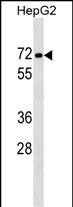
PPP1R16B Antibody (Center) 精选
Affinity Purified Rabbit Polyclonal Antibody (Pab)
- 产品详情
- 文献引用 : 4
- 实验流程
- 背景知识
Application 
| WB, E |
|---|---|
| Primary Accession | Q96T49 |
| Other Accession | Q8VHQ3, Q95N27, NP_001166206.1 |
| Reactivity | Human |
| Predicted | Bovine, Mouse, Rat, Canine, Rabbit |
| Host | Rabbit |
| Clonality | Polyclonal |
| Isotype | Rabbit IgG |
| Calculated MW | 63551 Da |
| Antigen Region | 372-399 aa |
| Gene ID | 26051 |
|---|---|
| Other Names | Protein phosphatase 1 regulatory inhibitor subunit 16B, Ankyrin repeat domain-containing protein 4, CAAX box protein TIMAP, TGF-beta-inhibited membrane-associated protein, hTIMAP, PPP1R16B, ANKRD4, KIAA0823 |
| Target/Specificity | This PPP1R16B antibody is generated from rabbits immunized with a KLH conjugated synthetic peptide between 372-399 amino acids from the Central region of human PPP1R16B. |
| Dilution | WB~~1:1000 E~~Use at an assay dependent concentration. |
| Format | Purified polyclonal antibody supplied in PBS with 0.09% (W/V) sodium azide. This antibody is purified through a protein A column, followed by peptide affinity purification. |
| Storage | Maintain refrigerated at 2-8°C for up to 2 weeks. For long term storage store at -20°C in small aliquots to prevent freeze-thaw cycles. |
| Precautions | PPP1R16B Antibody (Center) is for research use only and not for use in diagnostic or therapeutic procedures. |
For Research Use Only. Not For Use In Diagnostic Procedures.
| Name | PPP1R16B |
|---|---|
| Synonyms | ANKRD4, KIAA0823 |
| Function | Regulator of protein phosphatase 1 (PP1) that acts as a positive regulator of pulmonary endothelial cell (EC) barrier function (PubMed:18586956). Involved in the regulation of the PI3K/AKT signaling pathway, angiogenesis and endothelial cell proliferation (PubMed:25007873). Regulates angiogenesis and endothelial cell proliferation through the control of ECE1 dephosphorylation, trafficking and activity (By similarity). Protects the endothelial barrier from lipopolysaccharide (LPS)-induced vascular leakage (By similarity). Involved in the regulation of endothelial cell filopodia extension (By similarity). May be a downstream target for TGF-beta1 signaling cascade in endothelial cells (PubMed:16263087, PubMed:18586956). Involved in PKA-mediated moesin dephosphorylation which is important in EC barrier protection against thrombin stimulation (PubMed:18586956). Promotes the interaction of PPP1CA with RPSA/LAMR1 and in turn facilitates the dephosphorylation of RPSA/LAMR1 (PubMed:16263087). Involved in the dephosphorylation of EEF1A1 (PubMed:26497934). |
| Cellular Location | Cell membrane. Cell membrane; Lipid-anchor. Nucleus. Cell projection. Note=Colocalizes with RPSA/LAMR1 in the cell membrane (PubMed:16263087). Localizes to the perinuclear region (By similarity). Colocalizes with PTEN at the tip of EC projections (PubMed:25007873). {ECO:0000250|UniProtKB:Q95N27, ECO:0000269|PubMed:16263087, ECO:0000269|PubMed:25007873} |

Provided below are standard protocols that you may find useful for product applications.
BACKGROUND
The protein encoded by this gene is membrane-associated and contains five ankyrin repeats, a protein phosphatase-1-interacting domain, and a carboxy-terminal CAAX box domain. Synthesis of the encoded protein is inhibited by transforming growth factor beta-1. The protein may bind to the membrane through its CAAX box domain and may act as a signaling molecule through interaction with protein phosphatase-1. Alternatively spliced transcript variants encoding different isoforms have been identified in this gene.
REFERENCES
Csortos, C., et al. Am. J. Physiol. Lung Cell Mol. Physiol. 295 (3), L440-L450 (2008) :
Kim, K., et al. Biochem. Biophys. Res. Commun. 338(3):1327-1334(2005)
Homma, K., et al. J. Mol. Biol. 343(5):1207-1220(2004)
Cao, W., et al. Am. J. Physiol., Cell Physiol. 283 (1), C327-C337 (2002) :
Deloukas, P., et al. Nature 414(6866):865-871(2001)

终于等到您。ABCEPTA(百远生物)抗体产品。
点击下方“我要评价 ”按钮提交您的反馈信息,您的反馈和评价是我们最宝贵的财富之一,
我们将在1-3个工作日内处理您的反馈信息。
如有疑问,联系:0512-88856768 tech-china@abcepta.com.


