


 癌症的基本特征包括细胞增殖、血管生成、迁移、凋亡逃避机制和细胞永生等。找到癌症发生过程中这些通路的关键标记物和对应的抗体用于检测至关重要。
 为您推荐一个泛素化位点预测神器——泛素化分析工具,可以为您的蛋白的泛素化位点作出预测和评分。
 细胞自噬受体图形绘图工具为你的蛋白的细胞受体结合位点作出预测和评分,识别结合到自噬通路中的蛋白是非常重要的,便于让我们理解自噬在正常生理、病理过程中的作用,如发育、细胞分化、神经退化性疾病、压力条件下、感染和癌症。
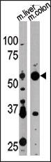
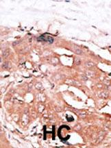
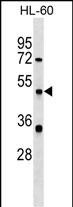
ATG4D Antibody (N-term)
Purified Rabbit Polyclonal Antibody (Pab)
- 产品详情
- 文献引用 : 2
- 实验流程
- 背景知识
Application
| WB, IHC-P, E |
|---|---|
| Primary Accession | Q86TL0 |
| Reactivity | Human, Mouse |
| Host | Rabbit |
| Clonality | Polyclonal |
| Isotype | Rabbit IgG |
| Calculated MW | 52922 Da |
| Antigen Region | 14-43 aa |
| Gene ID | 84971 |
|---|---|
| Other Names | Cysteine protease ATG4D, 3422-, AUT-like 4 cysteine endopeptidase, Autophagin-4, Autophagy-related cysteine endopeptidase 4, Autophagy-related protein 4 homolog D, Cysteine protease ATG4D, mitochondrial, ATG4D, APG4D, AUTL4 |
| Target/Specificity | This ATG4D antibody is generated from rabbits immunized with a KLH conjugated synthetic peptide between 14-43 amino acids from the N-terminal region of human ATG4D. |
| Dilution | WB~~1:1000 IHC-P~~1:100~500 E~~Use at an assay dependent concentration. |
| Format | Purified polyclonal antibody supplied in PBS with 0.09% (W/V) sodium azide. This antibody is prepared by Saturated Ammonium Sulfate (SAS) precipitation followed by dialysis against PBS. |
| Storage | Maintain refrigerated at 2-8°C for up to 2 weeks. For long term storage store at -20°C in small aliquots to prevent freeze-thaw cycles. |
| Precautions | ATG4D Antibody (N-term) is for research use only and not for use in diagnostic or therapeutic procedures. |
| Name | ATG4D {ECO:0000303|PubMed:19549685, ECO:0000312|HGNC:HGNC:20789} |
|---|---|
| Function | [Cysteine protease ATG4D]: Cysteine protease that plays a key role in autophagy by mediating both proteolytic activation and delipidation of ATG8 family proteins (PubMed:21177865, PubMed:29458288, PubMed:30661429). The protease activity is required for proteolytic activation of ATG8 family proteins: cleaves the C-terminal amino acid of ATG8 proteins MAP1LC3 and GABARAPL2, to reveal a C-terminal glycine (PubMed:21177865). Exposure of the glycine at the C-terminus is essential for ATG8 proteins conjugation to phosphatidylethanolamine (PE) and insertion to membranes, which is necessary for autophagy (By similarity). In addition to the protease activity, also mediates delipidation of ATG8 family proteins (PubMed:29458288, PubMed:33909989). Catalyzes delipidation of PE-conjugated forms of ATG8 proteins during macroautophagy (PubMed:29458288, PubMed:33909989). Also involved in non-canonical autophagy, a parallel pathway involving conjugation of ATG8 proteins to single membranes at endolysosomal compartments, by catalyzing delipidation of ATG8 proteins conjugated to phosphatidylserine (PS) (PubMed:33909989). ATG4D plays a role in the autophagy-mediated neuronal homeostasis in the central nervous system (By similarity). Compared to other members of the family (ATG4A, ATG4B or ATG4C), constitutes the major protein for the delipidation activity, while it promotes weak proteolytic activation of ATG8 proteins (By similarity). Involved in phagophore growth during mitophagy independently of its protease activity and of ATG8 proteins: acts by regulating ATG9A trafficking to mitochondria and promoting phagophore- endoplasmic reticulum contacts during the lipid transfer phase of mitophagy (PubMed:33773106). |
| Cellular Location | [Cysteine protease ATG4D]: Cytoplasm |
| Tissue Location | Widely expressed in testis. |
For Research Use Only. Not For Use In Diagnostic Procedures.

Provided below are standard protocols that you may find useful for product applications.
BACKGROUND
Macroautophagy is the major inducible pathway for the general turnover of cytoplasmic constituents in eukaryotic cells, it is also responsible for the degradation of active cytoplasmic enzymes and organelles during nutrient starvation. Macroautophagy involves the formation of double-membrane bound autophagosomes which enclose the cytoplasmic constituent targeted for degradation in a membrane bound structure, which then fuse with the lysosome (or vacuole) releasing a single-membrane bound autophagic bodies which are then degraded within the lysosome (or vacuole). APG4 is a cysteine protease required for autophagy, which cleaves the C-terminal part of either MAP1LC3, GABARAPL2 or GABARAP, allowing the liberation of form I. A subpopulation of form I is subsequently converted to a smaller form (form II). Form II, with a revealed C-terminal glycine, is considered to be the phosphatidylethanolamine (PE)-conjugated form, and has the capacity for the binding to autophagosomes.
REFERENCES
Baehrecke EH. Nat Rev Mol Cell Biol. 6(6):505-10. (2005) Lum JJ, et al. Nat Rev Mol Cell Biol. 6(6):439-48. (2005) Greenberg JT. Dev Cell. 8(6):799-801. (2005) Levine B. Cell. 120(2):159-62. (2005) Shintani T and Klionsky DJ. Science. 306(5698):990-5. (2004)
终于等到您。ABCEPTA(百远生物)抗体产品。
点击下方“我要评价 ”按钮提交您的反馈信息,您的反馈和评价是我们最宝贵的财富之一,
我们将在1-3个工作日内处理您的反馈信息。
如有疑问,联系:0512-88856768 tech-china@abcepta.com.


