


 癌症的基本特征包括细胞增殖、血管生成、迁移、凋亡逃避机制和细胞永生等。找到癌症发生过程中这些通路的关键标记物和对应的抗体用于检测至关重要。
 为您推荐一个泛素化位点预测神器——泛素化分析工具,可以为您的蛋白的泛素化位点作出预测和评分。
 细胞自噬受体图形绘图工具为你的蛋白的细胞受体结合位点作出预测和评分,识别结合到自噬通路中的蛋白是非常重要的,便于让我们理解自噬在正常生理、病理过程中的作用,如发育、细胞分化、神经退化性疾病、压力条件下、感染和癌症。
ATG5 Antibody (N-term)
Purified Rabbit Polyclonal Antibody (Pab)
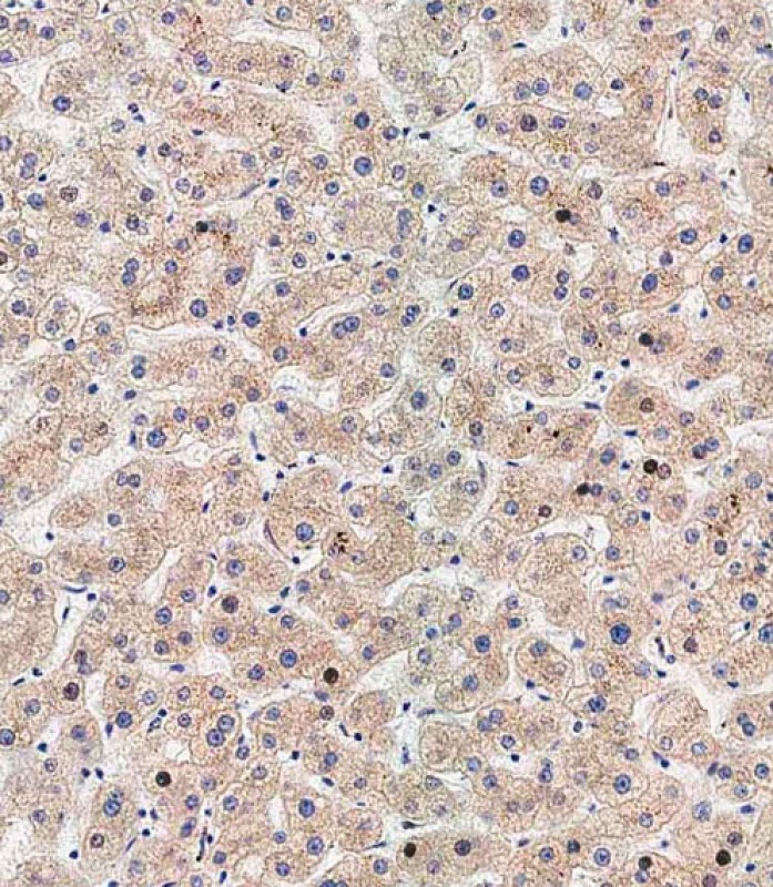
_-_U251_CQ.jpg)

- 产品详情
- 文献引用 : 28
- 实验流程
- 背景知识
Application
| WB, IF, IHC-P-Leica, E |
|---|---|
| Primary Accession | Q9H1Y0 |
| Other Accession | Q3MQ06, Q3MQ04, Q99J83, Q3MQ24 |
| Reactivity | Human, Rat, Mouse |
| Predicted | Mouse, Rat, Pig, Bovine |
| Host | Rabbit |
| Clonality | Polyclonal |
| Isotype | Rabbit IgG |
| Calculated MW | 32447 Da |
| Antigen Region | 1-30 aa |
| Gene ID | 9474 |
|---|---|
| Other Names | Autophagy protein 5, APG5-like, Apoptosis-specific protein, ATG5, APG5L, ASP |
| Target/Specificity | This ATG5 antibody is generated from rabbits immunized with a KLH conjugated synthetic peptide between 1-30 amino acids from the N-terminal region of human ATG5. |
| Dilution | WB~~1:1000 IF~~1:200 IHC-P-Leica~~1:500 E~~Use at an assay dependent concentration. |
| Format | Purified polyclonal antibody supplied in PBS with 0.09% (W/V) sodium azide. This antibody is purified through a protein A column, followed by peptide affinity purification. |
| Storage | Maintain refrigerated at 2-8°C for up to 2 weeks. For long term storage store at -20°C in small aliquots to prevent freeze-thaw cycles. |
| Precautions | ATG5 Antibody (N-term) is for research use only and not for use in diagnostic or therapeutic procedures. |
| Name | ATG5 (HGNC:589) |
|---|---|
| Synonyms | APG5L, ASP |
| Function | Involved in autophagic vesicle formation. Conjugation with ATG12, through a ubiquitin-like conjugating system involving ATG7 as an E1-like activating enzyme and ATG10 as an E2-like conjugating enzyme, is essential for its function. The ATG12-ATG5 conjugate acts as an E3- like enzyme which is required for lipidation of ATG8 family proteins and their association to the vesicle membranes. Involved in mitochondrial quality control after oxidative damage, and in subsequent cellular longevity. Plays a critical role in multiple aspects of lymphocyte development and is essential for both B and T lymphocyte survival and proliferation. Required for optimal processing and presentation of antigens for MHC II. Involved in the maintenance of axon morphology and membrane structures, as well as in normal adipocyte differentiation. Promotes primary ciliogenesis through removal of OFD1 from centriolar satellites and degradation of IFT20 via the autophagic pathway. As part of the ATG8 conjugation system with ATG12 and ATG16L1, required for recruitment of LRRK2 to stressed lysosomes and induction of LRRK2 kinase activity in response to lysosomal stress (By similarity). |
| Cellular Location | Cytoplasm. Preautophagosomal structure membrane; Peripheral membrane protein. Note=Colocalizes with nonmuscle actin. The conjugate detaches from the membrane immediately before or after autophagosome formation is completed (By similarity). Also localizes to discrete punctae along the ciliary axoneme and to the base of the ciliary axoneme. Under starved conditions, the ATG12-ATG5-ATG16L1 complex is translocated to phagophores driven by RAB33B (PubMed:32960676). {ECO:0000250, ECO:0000269|PubMed:32960676} |
| Tissue Location | Ubiquitous. The mRNA is present at similar levels in viable and apoptotic cells, whereas the protein is dramatically highly expressed in apoptotic cells |
For Research Use Only. Not For Use In Diagnostic Procedures.

Provided below are standard protocols that you may find useful for product applications.
BACKGROUND
Macroautophagy is the major inducible pathway for the general turnover of cytoplasmic constituents in eukaryotic cells, it is also responsible for the degradation of active cytoplasmic enzymes and organelles during nutrient starvation. Macroautophagy involves the formation of double-membrane bound autophagosomes which enclose the cytoplasmic constituent targeted for degradation in a membrane bound structure, which then fuse with the lysosome (or vacuole) releasing a single-membrane bound autophagic bodies which are then degraded within the lysosome (or vacuole). APG5, required for autophagy, conjugates to ATG12 and associates with an isolation membrane to form a cup-shaped isolation membrane and autophagosome. The conjugate detaches from the membrane immediately before or after autophagosome formation is completed. APG5 may also play an important role in the apoptotic process, possibly within the modified cytoskeleton. Its expression is a relatively late event in the apoptotic process, occurring downstream of caspase activity.
REFERENCES
References for protein:
1.Baehrecke EH. Nat Rev Mol Cell Biol. 6(6):505-10. (2005)
2. Lum JJ, et al. Nat Rev Mol Cell Biol. 6(6):439-48. (2005)
3.Greenberg JT. Dev Cell. 8(6):799-801. (2005)
4.Levine B. Cell. 120(2):159-62. (2005)
5.Shintani T and Klionsky DJ. Science. 306(5698):990-5. (2004)
6.Hammond E.M., et al. FEBS Lett. 425:391-395(1998)
7. Strausberg R.L., et al. PNAS 99:16899-16903(2002)
8.Grand R.J.A., et al. Exp. Cell Res. 218:439-451(1995)
9.Mizushima N., et al. J. Biol. Chem. 273:33889-33892(1998)
10.Mizushima N., et al. J. Cell Biol. 152:657-668(2001)
References for U251 cell line:
1. Westermark B.; Pontén J.; Hugosson R. (1973).” Determinants for the establishment of permanent tissue culture lines from human gliomas”. Acta Pathol Microbiol Scand A. 81:791-805. [PMID: 4359449].
2. Pontén, J.,Westermark B. (1978).” Properties of Human Malignant Glioma Cells in Vitro”. Medical Biology 56: 184-193.[PMID: 359950].
3. Geng Y.;Kohli L.; Klocke B.J.; Roth K.A.(2010). “Chloroquine-induced autophagic vacuole accumulation and cell death in glioma cells is p53 independent”. Neuro Oncol. 12(5): 473–481.[ PMID: 20406898].
终于等到您。ABCEPTA(百远生物)抗体产品。
点击下方“我要评价 ”按钮提交您的反馈信息,您的反馈和评价是我们最宝贵的财富之一,
我们将在1-3个工作日内处理您的反馈信息。
如有疑问,联系:0512-88856768 tech-china@abcepta.com.


