


 癌症的基本特征包括细胞增殖、血管生成、迁移、凋亡逃避机制和细胞永生等。找到癌症发生过程中这些通路的关键标记物和对应的抗体用于检测至关重要。
 为您推荐一个泛素化位点预测神器——泛素化分析工具,可以为您的蛋白的泛素化位点作出预测和评分。
 细胞自噬受体图形绘图工具为你的蛋白的细胞受体结合位点作出预测和评分,识别结合到自噬通路中的蛋白是非常重要的,便于让我们理解自噬在正常生理、病理过程中的作用,如发育、细胞分化、神经退化性疾病、压力条件下、感染和癌症。
Anti-MK2 (pT222) Antibody
Rabbit polyclonal antibody to MK2 (pT222)
- 产品详情
- 实验流程
- 背景知识
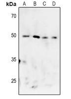
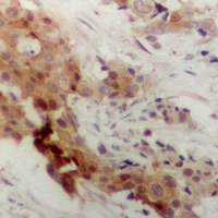
Application
| WB, IHC |
|---|---|
| Primary Accession | P49137 |
| Other Accession | P49138 |
| Reactivity | Human, Mouse, Rat, Bovine |
| Host | Rabbit |
| Clonality | Polyclonal |
| Calculated MW | 45568 Da |
| Gene ID | 9261 |
|---|---|
| Other Names | MAP kinase-activated protein kinase 2; MAPK-activated protein kinase 2; MAPKAP kinase 2; MAPKAP-K2; MAPKAPK-2; MK-2; MK2 |
| Target/Specificity | KLH-conjugated synthetic peptide encompassing a sequence within the center region of human MK2. The exact sequence is proprietary. |
| Dilution | WB~~WB (1/500 - 1/1000), IHC (1/50 - 1/100) IHC~~WB (1/500 - 1/1000), IHC (1/50 - 1/100) |
| Format | Liquid in 0.42% Potassium phosphate, 0.87% Sodium chloride, pH 7.3, 30% glycerol, and 0.09% (W/V) sodium azide. |
| Storage | Store at -20 °C.Stable for 12 months from date of receipt |
| Name | MAPKAPK2 |
|---|---|
| Function | Stress-activated serine/threonine-protein kinase involved in cytokine production, endocytosis, reorganization of the cytoskeleton, cell migration, cell cycle control, chromatin remodeling, DNA damage response and transcriptional regulation. Following stress, it is phosphorylated and activated by MAP kinase p38-alpha/MAPK14, leading to phosphorylation of substrates. Phosphorylates serine in the peptide sequence, Hyd-X-R-X(2)-S, where Hyd is a large hydrophobic residue. Phosphorylates ALOX5, CDC25B, CDC25C, CEP131, ELAVL1, HNRNPA0, HSP27/HSPB1, KRT18, KRT20, LIMK1, LSP1, PABPC1, PARN, PDE4A, RCSD1, RPS6KA3, TAB3 and TTP/ZFP36. Phosphorylates HSF1; leading to the interaction with HSP90 proteins and inhibiting HSF1 homotrimerization, DNA-binding and transactivation activities (PubMed:16278218). Mediates phosphorylation of HSP27/HSPB1 in response to stress, leading to the dissociation of HSP27/HSPB1 from large small heat-shock protein (sHsps) oligomers and impairment of their chaperone activities and ability to protect against oxidative stress effectively. Involved in inflammatory response by regulating tumor necrosis factor (TNF) and IL6 production post-transcriptionally: acts by phosphorylating AU-rich elements (AREs)-binding proteins ELAVL1, HNRNPA0, PABPC1 and TTP/ZFP36, leading to the regulation of the stability and translation of TNF and IL6 mRNAs. Phosphorylation of TTP/ZFP36, a major post-transcriptional regulator of TNF, promotes its binding to 14-3-3 proteins and reduces its ARE mRNA affinity, leading to inhibition of dependent degradation of ARE-containing transcripts. Phosphorylates CEP131 in response to cellular stress induced by ultraviolet irradiation which promotes binding of CEP131 to 14-3-3 proteins and inhibits formation of novel centriolar satellites (PubMed:26616734). Also involved in late G2/M checkpoint following DNA damage through a process of post- transcriptional mRNA stabilization: following DNA damage, relocalizes from nucleus to cytoplasm and phosphorylates HNRNPA0 and PARN, leading to stabilization of GADD45A mRNA. Involved in toll-like receptor signaling pathway (TLR) in dendritic cells: required for acute TLR- induced macropinocytosis by phosphorylating and activating RPS6KA3. |
| Cellular Location | Cytoplasm. Nucleus. Note=Phosphorylation and subsequent activation releases the autoinhibitory helix, resulting in the export from the nucleus into the cytoplasm |
| Tissue Location | Expressed in all tissues examined. |
Research Areas
For Research Use Only. Not For Use In Diagnostic Procedures.
Application Protocols
Provided below are standard protocols that you may find useful for product applications.
BACKGROUND
KLH-conjugated synthetic peptide encompassing a sequence within the center region of human MK2. The exact sequence is proprietary.
终于等到您。ABCEPTA(百远生物)抗体产品。
点击下方“我要评价 ”按钮提交您的反馈信息,您的反馈和评价是我们最宝贵的财富之一,
我们将在1-3个工作日内处理您的反馈信息。
如有疑问,联系:0512-88856768 tech-china@abcepta.com.
¥ 1,500.00
Cat# AP61068


