


 癌症的基本特征包括细胞增殖、血管生成、迁移、凋亡逃避机制和细胞永生等。找到癌症发生过程中这些通路的关键标记物和对应的抗体用于检测至关重要。
 为您推荐一个泛素化位点预测神器——泛素化分析工具,可以为您的蛋白的泛素化位点作出预测和评分。
 细胞自噬受体图形绘图工具为你的蛋白的细胞受体结合位点作出预测和评分,识别结合到自噬通路中的蛋白是非常重要的,便于让我们理解自噬在正常生理、病理过程中的作用,如发育、细胞分化、神经退化性疾病、压力条件下、感染和癌症。
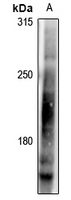
Anti-LRP6 (pT1479) Antibody
Rabbit polyclonal antibody to LRP6 (pT1479)
- 产品详情
- 实验流程
- 背景知识
Application
| WB |
|---|---|
| Primary Accession | O75581 |
| Other Accession | O88572 |
| Reactivity | Human, Mouse |
| Host | Rabbit |
| Clonality | Polyclonal |
| Calculated MW | 180429 Da |
| Gene ID | 4040 |
|---|---|
| Other Names | Low-density lipoprotein receptor-related protein 6; LRP-6 |
| Target/Specificity | KLH-conjugated synthetic peptide encompassing a sequence within the C-term region of human LRP6 (pT1479). The exact sequence is proprietary. |
| Dilution | WB~~WB (1/500 - 1/1000) |
| Format | Liquid in 0.42% Potassium phosphate, 0.87% Sodium chloride, pH 7.3, 30% glycerol, and 0.09% (W/V) sodium azide. |
| Storage | Store at -20 °C.Stable for 12 months from date of receipt |
| Name | LRP6 |
|---|---|
| Function | Component of the Wnt-Fzd-LRP5-LRP6 complex that triggers beta-catenin signaling through inducing aggregation of receptor-ligand complexes into ribosome-sized signalosomes (PubMed:11357136, PubMed:11448771, PubMed:15778503, PubMed:16341017, PubMed:16513652, PubMed:17326769, PubMed:17400545, PubMed:19107203, PubMed:19293931, PubMed:19801552, PubMed:28341812). Cell-surface coreceptor of Wnt/beta- catenin signaling, which plays a pivotal role in bone formation (PubMed:11357136, PubMed:11448771, PubMed:15778503, PubMed:16341017, PubMed:16513652, PubMed:17326769, PubMed:17400545, PubMed:19107203, PubMed:19293931, PubMed:19801552, PubMed:28341812). The Wnt-induced Fzd/LRP6 coreceptor complex recruits DVL1 polymers to the plasma membrane which, in turn, recruits the AXIN1/GSK3B-complex to the cell surface promoting the formation of signalosomes and inhibiting AXIN1/GSK3-mediated phosphorylation and destruction of beta-catenin (PubMed:16513652). Required for posterior patterning of the epiblast during gastrulation (By similarity). |
| Cellular Location | Cell membrane; Single-pass type I membrane protein Endoplasmic reticulum. Membrane raft. Note=On Wnt signaling, undergoes a cycle of caveolin- or clathrin-mediated endocytosis and plasma membrane location. Released from the endoplasmic reticulum on palmitoylation Mono-ubiquitination retains it in the endoplasmic reticulum in the absence of palmitoylation. On Wnt signaling, phosphorylated, aggregates and colocalizes with AXIN1 and GSK3B at the plasma membrane in LRP6- signalosomes (By similarity). Chaperoned to the plasma membrane by HSP90B1 and MESD (PubMed:23572575). {ECO:0000250|UniProtKB:O88572, ECO:0000269|PubMed:23572575} |
| Tissue Location | Widely coexpressed with LRP5 during embryogenesis and in adult tissues |
Research Areas
For Research Use Only. Not For Use In Diagnostic Procedures.
Application Protocols
Provided below are standard protocols that you may find useful for product applications.
BACKGROUND
KLH-conjugated synthetic peptide encompassing a sequence within the C-term region of human LRP6 (pT1479). The exact sequence is proprietary.
终于等到您。ABCEPTA(百远生物)抗体产品。
点击下方“我要评价 ”按钮提交您的反馈信息,您的反馈和评价是我们最宝贵的财富之一,
我们将在1-3个工作日内处理您的反馈信息。
如有疑问,联系:0512-88856768 tech-china@abcepta.com.
¥ 1,500.00
Cat# AP61279


