


 癌症的基本特征包括细胞增殖、血管生成、迁移、凋亡逃避机制和细胞永生等。找到癌症发生过程中这些通路的关键标记物和对应的抗体用于检测至关重要。
 为您推荐一个泛素化位点预测神器——泛素化分析工具,可以为您的蛋白的泛素化位点作出预测和评分。
 细胞自噬受体图形绘图工具为你的蛋白的细胞受体结合位点作出预测和评分,识别结合到自噬通路中的蛋白是非常重要的,便于让我们理解自噬在正常生理、病理过程中的作用,如发育、细胞分化、神经退化性疾病、压力条件下、感染和癌症。
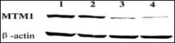
MTM1 Antibody (C-term)
Purified Rabbit Polyclonal Antibody (Pab)
- 产品详情
- 文献引用 : 1
- 实验流程
- 背景知识
Application
| WB, E |
|---|---|
| Primary Accession | Q13496 |
| Reactivity | Human, Mouse |
| Host | Rabbit |
| Clonality | Polyclonal |
| Isotype | Rabbit IgG |
| Calculated MW | 69932 Da |
| Antigen Region | 566-594 aa |
| Gene ID | 4534 |
|---|---|
| Other Names | Myotubularin, Phosphatidylinositol-3, 5-bisphosphate 3-phosphatase, Phosphatidylinositol-3-phosphate phosphatase, MTM1, CG2 |
| Target/Specificity | This MTM1 antibody is generated from rabbits immunized with a KLH conjugated synthetic peptide between 566-594 amino acids from the C-terminal region of human MTM1. |
| Dilution | WB~~1:1000 E~~Use at an assay dependent concentration. |
| Format | Purified polyclonal antibody supplied in PBS with 0.09% (W/V) sodium azide. This antibody is prepared by Saturated Ammonium Sulfate (SAS) precipitation followed by dialysis against PBS. |
| Storage | Maintain refrigerated at 2-8°C for up to 2 weeks. For long term storage store at -20°C in small aliquots to prevent freeze-thaw cycles. |
| Precautions | MTM1 Antibody (C-term) is for research use only and not for use in diagnostic or therapeutic procedures. |
| Name | MTM1 (HGNC:7448) |
|---|---|
| Synonyms | CG2 |
| Function | Lipid phosphatase which dephosphorylates phosphatidylinositol 3-monophosphate (PI3P) and phosphatidylinositol 3,5-bisphosphate (PI(3,5)P2) (PubMed:10900271, PubMed:11001925, PubMed:12646134, PubMed:14722070). Has also been shown to dephosphorylate phosphotyrosine- and phosphoserine-containing peptides (PubMed:9537414). Negatively regulates EGFR degradation through regulation of EGFR trafficking from the late endosome to the lysosome (PubMed:14722070). Plays a role in vacuolar formation and morphology. Regulates desmin intermediate filament assembly and architecture (PubMed:21135508). Plays a role in mitochondrial morphology and positioning (PubMed:21135508). Required for skeletal muscle maintenance but not for myogenesis (PubMed:21135508). In skeletal muscles, stabilizes MTMR12 protein levels (PubMed:23818870). |
| Cellular Location | Cytoplasm. Cell membrane; Peripheral membrane protein. Cell projection, filopodium. Cell projection, ruffle. Late endosome. Cytoplasm, myofibril, sarcomere {ECO:0000250|UniProtKB:Q9Z2C5}. Note=Localizes as a dense cytoplasmic network (PubMed:11001925). Also localizes to the plasma membrane, including plasma membrane extensions such as filopodia and ruffles (PubMed:12118066). Predominantly located in the cytoplasm following interaction with MTMR12 (PubMed:12847286). Recruited to the late endosome following EGF stimulation (PubMed:14722070). In skeletal muscles, co-localizes with MTMR12 in the sarcomere (By similarity) {ECO:0000250|UniProtKB:Q9Z2C5, ECO:0000269|PubMed:11001925, ECO:0000269|PubMed:12118066, ECO:0000269|PubMed:12847286, ECO:0000269|PubMed:14722070} |
For Research Use Only. Not For Use In Diagnostic Procedures.

Provided below are standard protocols that you may find useful for product applications.
BACKGROUND
MTM1 is a member of a protein family that encodes tyrosine phosphatases. Myotubularin is required for muscle cell differentiation and mutations in MTM1 have been identified as being responsible for X-linked myotubular myopathy. MTM1 is a potent phosphatidylinositol 3-phosphate phosphatase (PI(3)P). Mutations in the MTM1 gene that cause human myotubular myopathy dramatically reduce the ability of the phosphatase to dephosphorylate PI(3)P. The findings provided evidence that myotubularin exerts its effects during myogenesis by regulating the cellular levels of the inositol lipid PI(3)P.
REFERENCES
Nandurkar, H.H., et al., Proc. Natl. Acad. Sci. U.S.A. 100(15):8660-8665 (2003).
Biancalana, V., et al., Hum. Genet. 112(2):135-142 (2003).
Wishart, M.J., et al., Trends Cell Biol. 12(12):579-585 (2002).
Herman, G.E., et al., Hum. Mutat. 19(2):114-121 (2002).
Sutton, I.J., et al., Neurology 57(5):900-902 (2001).
终于等到您。ABCEPTA(百远生物)抗体产品。
点击下方“我要评价 ”按钮提交您的反馈信息,您的反馈和评价是我们最宝贵的财富之一,
我们将在1-3个工作日内处理您的反馈信息。
如有疑问,联系:0512-88856768 tech-china@abcepta.com.


