


 癌症的基本特征包括细胞增殖、血管生成、迁移、凋亡逃避机制和细胞永生等。找到癌症发生过程中这些通路的关键标记物和对应的抗体用于检测至关重要。
 为您推荐一个泛素化位点预测神器——泛素化分析工具,可以为您的蛋白的泛素化位点作出预测和评分。
 细胞自噬受体图形绘图工具为你的蛋白的细胞受体结合位点作出预测和评分,识别结合到自噬通路中的蛋白是非常重要的,便于让我们理解自噬在正常生理、病理过程中的作用,如发育、细胞分化、神经退化性疾病、压力条件下、感染和癌症。
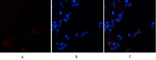
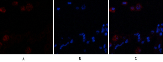
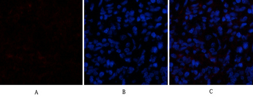
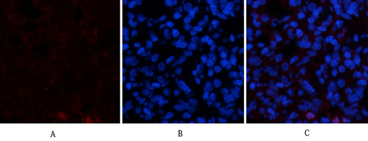
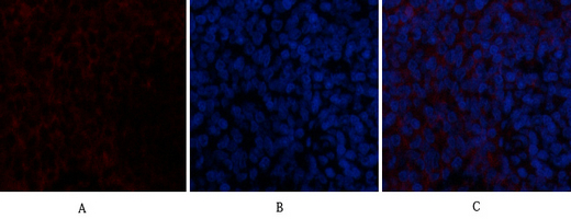
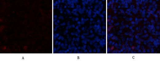
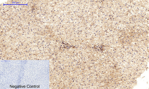
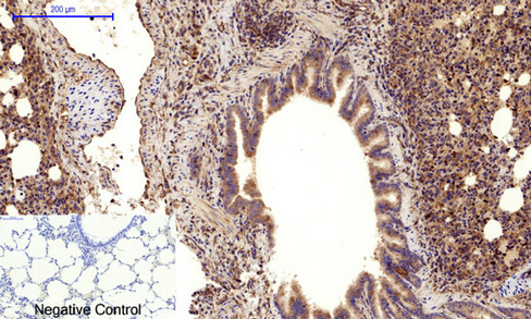
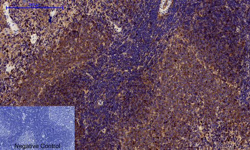
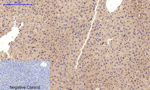
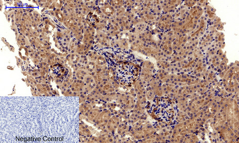
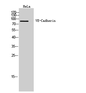
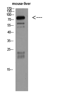
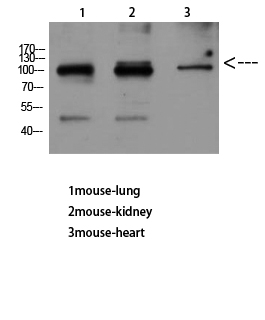
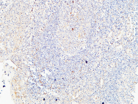
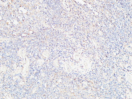
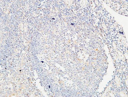
VE-Cadherin Polyclonal Antibody
- 产品详情
- 文献引用 : 1
- 实验流程
- 背景知识
Application
| WB, IHC-P, IF |
|---|---|
| Primary Accession | P33151 |
| Reactivity | Human, Mouse, Rat |
| Host | Rabbit |
| Clonality | Polyclonal |
| Calculated MW | 87528 Da |
| Gene ID | 1003 |
|---|---|
| Other Names | CDH5; Cadherin-5; 7B4 antigen; Vascular endothelial cadherin; VE-cadherin; CD144 |
| Dilution | WB~~IF: 1:50-200 WB 1:500-2000, ELISA 1:10000-20000 IHC 1:50-300 IHC-P~~IF: 1:50-200 WB 1:500-2000, ELISA 1:10000-20000 IHC 1:50-300 IF~~IF: 1:50-200 WB 1:500-2000, ELISA 1:10000-20000 IHC 1:50-300 |
| Format | Liquid in PBS containing 50% glycerol, 0.5% BSA and 0.09% (W/V) sodium azide. |
| Storage Conditions | -20℃ |
| Name | CDH5 (HGNC:1764) |
|---|---|
| Function | Cadherins are calcium-dependent cell adhesion proteins (By similarity). They preferentially interact with themselves in a homophilic manner in connecting cells; cadherins may thus contribute to the sorting of heterogeneous cell types (PubMed:21269602). This cadherin may play a important role in endothelial cell biology through control of the cohesion and organization of the intercellular junctions (By similarity). It associates with alpha-catenin forming a link to the cytoskeleton (PubMed:10861224). Plays a role in coupling actin fibers to cell junctions in endothelial cells, via acting as a cell junctional complex anchor for AMOTL2 and MAGI1 (By similarity). Acts in concert with KRIT1 and PALS1 to establish and maintain correct endothelial cell polarity and vascular lumen (By similarity). These effects are mediated by recruitment and activation of the Par polarity complex and RAP1B (PubMed:20332120). Required for activation of PRKCZ and for the localization of phosphorylated PRKCZ, PARD3, TIAM1 and RAP1B to the cell junction (PubMed:20332120). Associates with CTNND1/p120-catenin to control CADH5 endocytosis (By similarity). |
| Cellular Location | Cell junction, adherens junction. Cell membrane; Single-pass type I membrane protein. Cytoplasm {ECO:0000250|UniProtKB:P55284}. Note=Found at cell-cell boundaries and probably at cell-matrix boundaries. KRIT1 and CDH5 reciprocally regulate their localization to endothelial cell-cell junctions |
| Tissue Location | Expressed in endothelial cells (at protein level) (PubMed:27338829). Expressed in the brain (PubMed:2059658) |
For Research Use Only. Not For Use In Diagnostic Procedures.

Provided below are standard protocols that you may find useful for product applications.
BACKGROUND
Cadherins are calcium-dependent cell adhesion proteins. They preferentially interact with themselves in a homophilic manner in connecting cells; cadherins may thus contribute to the sorting of heterogeneous cell types. This cadherin may play a important role in endothelial cell biology through control of the cohesion and organization of the intercellular junctions. It associates with alpha-catenin forming a link to the cytoskeleton. Acts in concert with KRIT1 to establish and maintain correct endothelial cell polarity and vascular lumen. These effects are mediated by recruitment and activation of the Par polarity complex and RAP1B. Required for activation of PRKCZ and for the localization of phosphorylated PRKCZ, PARD3, TIAM1 and RAP1B to the cell junction.
终于等到您。ABCEPTA(百远生物)抗体产品。
点击下方“我要评价 ”按钮提交您的反馈信息,您的反馈和评价是我们最宝贵的财富之一,
我们将在1-3个工作日内处理您的反馈信息。
如有疑问,联系:0512-88856768 tech-china@abcepta.com.


