


 癌症的基本特征包括细胞增殖、血管生成、迁移、凋亡逃避机制和细胞永生等。找到癌症发生过程中这些通路的关键标记物和对应的抗体用于检测至关重要。
 为您推荐一个泛素化位点预测神器——泛素化分析工具,可以为您的蛋白的泛素化位点作出预测和评分。
 细胞自噬受体图形绘图工具为你的蛋白的细胞受体结合位点作出预测和评分,识别结合到自噬通路中的蛋白是非常重要的,便于让我们理解自噬在正常生理、病理过程中的作用,如发育、细胞分化、神经退化性疾病、压力条件下、感染和癌症。
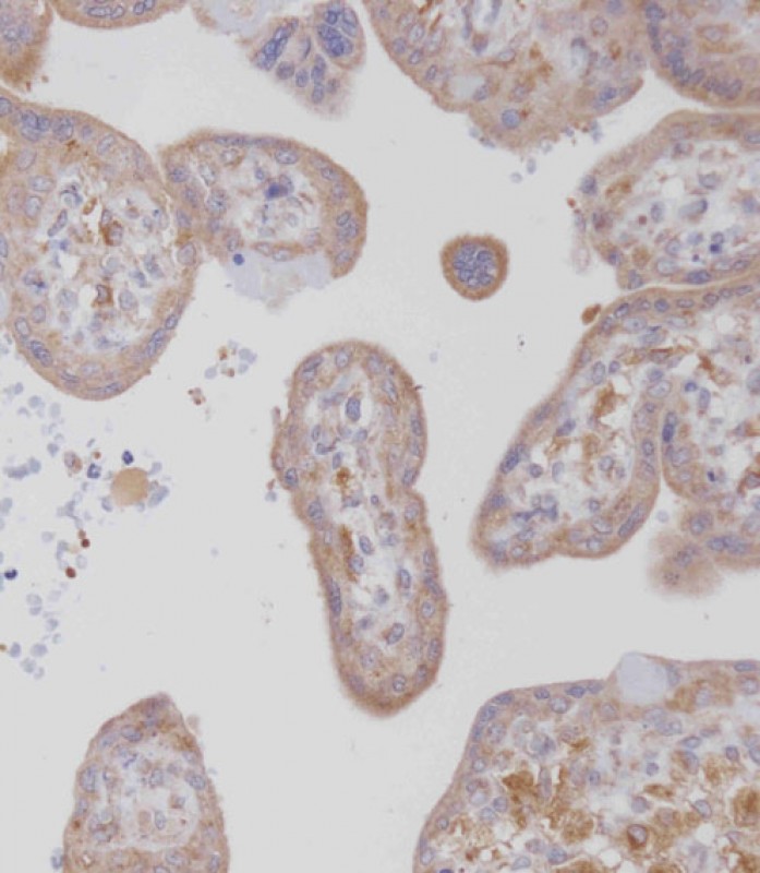
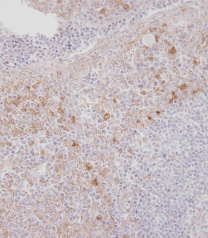
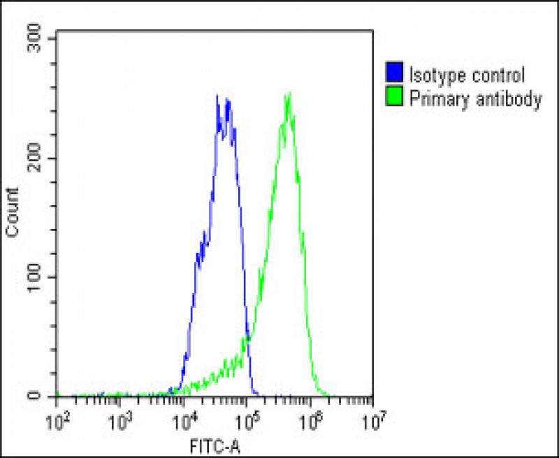
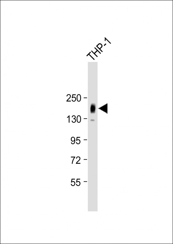
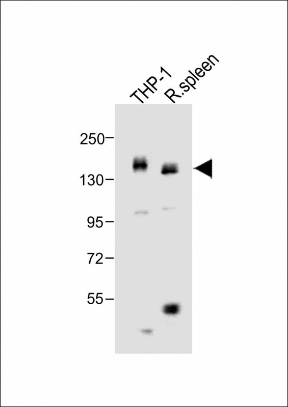
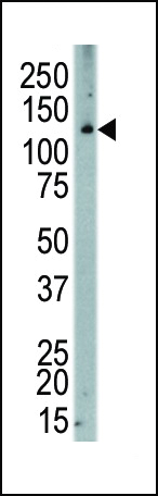
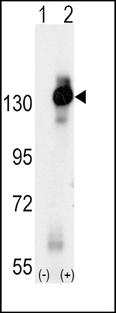
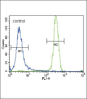
MCSF Receptor (CSF1R) Antibody (C-term)
Purified Rabbit Polyclonal Antibody (Pab)
- 产品详情
- 文献引用 : 1
- 实验流程
- 背景知识
Application
| IHC-P, WB, FC, E |
|---|---|
| Primary Accession | P07333 |
| Reactivity | Human, Rat, Mouse |
| Host | Rabbit |
| Clonality | Polyclonal |
| Isotype | Rabbit IgG |
| Calculated MW | 107984 Da |
| Antigen Region | 940-971 aa |
| Gene ID | 1436 |
|---|---|
| Other Names | Macrophage colony-stimulating factor 1 receptor, CSF-1 receptor, CSF-1-R, CSF-1R, M-CSF-R, Proto-oncogene c-Fms, CD115, CSF1R, FMS |
| Target/Specificity | This MCSF Receptor (CSF1R) antibody is generated from rabbits immunized with a KLH conjugated synthetic peptide between 940-971 amino acids from the C-terminal region of human MCSF Receptor (CSF1R). |
| Dilution | IHC-P~~1:100~500 WB~~1:1000 FC~~1:10~50 E~~Use at an assay dependent concentration. |
| Format | Purified polyclonal antibody supplied in PBS with 0.09% (W/V) sodium azide. This antibody is purified through a protein A column, followed by peptide affinity purification. |
| Storage | Maintain refrigerated at 2-8°C for up to 2 weeks. For long term storage store at -20°C in small aliquots to prevent freeze-thaw cycles. |
| Precautions | MCSF Receptor (CSF1R) Antibody (C-term) is for research use only and not for use in diagnostic or therapeutic procedures. |
| Name | CSF1R |
|---|---|
| Synonyms | FMS |
| Function | Tyrosine-protein kinase that acts as a cell-surface receptor for CSF1 and IL34 and plays an essential role in the regulation of survival, proliferation and differentiation of hematopoietic precursor cells, especially mononuclear phagocytes, such as macrophages and monocytes. Promotes the release of pro-inflammatory chemokines in response to IL34 and CSF1, and thereby plays an important role in innate immunity and in inflammatory processes. Plays an important role in the regulation of osteoclast proliferation and differentiation, the regulation of bone resorption, and is required for normal bone and tooth development. Required for normal male and female fertility, and for normal development of milk ducts and acinar structures in the mammary gland during pregnancy. Promotes reorganization of the actin cytoskeleton, regulates formation of membrane ruffles, cell adhesion and cell migration, and promotes cancer cell invasion. Activates several signaling pathways in response to ligand binding, including the ERK1/2 and the JNK pathway (PubMed:20504948, PubMed:30982609). Phosphorylates PIK3R1, PLCG2, GRB2, SLA2 and CBL. Activation of PLCG2 leads to the production of the cellular signaling molecules diacylglycerol and inositol 1,4,5-trisphosphate, that then lead to the activation of protein kinase C family members, especially PRKCD. Phosphorylation of PIK3R1, the regulatory subunit of phosphatidylinositol 3-kinase, leads to activation of the AKT1 signaling pathway. Activated CSF1R also mediates activation of the MAP kinases MAPK1/ERK2 and/or MAPK3/ERK1, and of the SRC family kinases SRC, FYN and YES1. Activated CSF1R transmits signals both via proteins that directly interact with phosphorylated tyrosine residues in its intracellular domain, or via adapter proteins, such as GRB2. Promotes activation of STAT family members STAT3, STAT5A and/or STAT5B. Promotes tyrosine phosphorylation of SHC1 and INPP5D/SHIP-1. Receptor signaling is down-regulated by protein phosphatases, such as INPP5D/SHIP-1, that dephosphorylate the receptor and its downstream effectors, and by rapid internalization of the activated receptor. In the central nervous system, may play a role in the development of microglia macrophages (PubMed:30982608). |
| Cellular Location | Cell membrane; Single-pass type I membrane protein |
| Tissue Location | Expressed in bone marrow and in differentiated blood mononuclear cells |
For Research Use Only. Not For Use In Diagnostic Procedures.

Provided below are standard protocols that you may find useful for product applications.
BACKGROUND
CSF1R is the receptor for colony stimulating factor 1, a cytokine which controls the production, differentiation, and function of macrophages. This receptor mediates most if not all of the biological effects of this cytokine. Ligand binding activates the receptor kinase through a process of oligomerization and transphosphorylation. This protein is a tyrosine kinase transmembrane receptor and member of the CSF1/PDGF receptor family of tyrosine-protein kinases. Mutations in the gene encoding CSF1R have been associated with a predisposition to myeloid malignancy.
REFERENCES
Follows, G.A., et al., EMBO J. 22(11):2798-2809 (2003).
Riccioni, R., et al., Leukemia 17(1):98-113 (2003).
Zhu, K., et al., Biochem. Biophys. Res. Commun. 297(5):1211-1217 (2002).
Ide, H., et al., Proc. Natl. Acad. Sci. U.S.A. 99(22):14404-14409 (2002).
Flick, M.B., et al., J. Cell. Biochem. 85(1):10-23 (2002).
终于等到您。ABCEPTA(百远生物)抗体产品。
点击下方“我要评价 ”按钮提交您的反馈信息,您的反馈和评价是我们最宝贵的财富之一,
我们将在1-3个工作日内处理您的反馈信息。
如有疑问,联系:0512-88856768 tech-china@abcepta.com.


