


 癌症的基本特征包括细胞增殖、血管生成、迁移、凋亡逃避机制和细胞永生等。找到癌症发生过程中这些通路的关键标记物和对应的抗体用于检测至关重要。
 为您推荐一个泛素化位点预测神器——泛素化分析工具,可以为您的蛋白的泛素化位点作出预测和评分。
 细胞自噬受体图形绘图工具为你的蛋白的细胞受体结合位点作出预测和评分,识别结合到自噬通路中的蛋白是非常重要的,便于让我们理解自噬在正常生理、病理过程中的作用,如发育、细胞分化、神经退化性疾病、压力条件下、感染和癌症。
BAI1 Antibody (C-term)
Affinity Purified Rabbit Polyclonal Antibody (Pab)
- 产品详情
- 文献引用 : 1
- 实验流程
- 背景知识
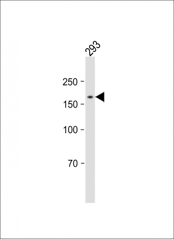
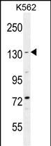
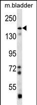
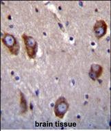
Application
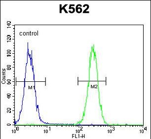
| WB, IHC-P, FC, E |
|---|---|
| Primary Accession | O14514 |
| Reactivity | Human, Mouse |
| Host | Rabbit |
| Clonality | Polyclonal |
| Isotype | Rabbit IgG |
| Calculated MW | 173501 Da |
| Antigen Region | 1537-1567 aa |
| Gene ID | 575 |
|---|---|
| Other Names | Brain-specific angiogenesis inhibitor 1, BAI1 |
| Target/Specificity | This BAI1 antibody is generated from rabbits immunized with a KLH conjugated synthetic peptide between 1537-1567 amino acids from the C-terminal region of human BAI1. |
| Dilution | WB~~1:1000 IHC-P~~1:100~500 FC~~1:10~50 E~~Use at an assay dependent concentration. |
| Format | Purified polyclonal antibody supplied in PBS with 0.05% (V/V) Proclin 300. This antibody is purified through a protein A column, followed by peptide affinity purification. |
| Storage | Maintain refrigerated at 2-8°C for up to 2 weeks. For long term storage store at -20°C in small aliquots to prevent freeze-thaw cycles. |
| Precautions | BAI1 Antibody (C-term) is for research use only and not for use in diagnostic or therapeutic procedures. |
| Name | ADGRB1 (HGNC:943) |
|---|---|
| Function | Phosphatidylserine receptor which enhances the engulfment of apoptotic cells (PubMed:24509909). Also mediates the binding and engulfment of Gram-negative bacteria (PubMed:26838550). Stimulates production of reactive oxygen species by macrophages in response to Gram-negative bacteria, resulting in enhanced microbicidal macrophage activity (PubMed:26838550). In the gastric mucosa, required for recognition and engulfment of apoptotic gastric epithelial cells (PubMed:24509909). Promotes myoblast fusion (By similarity). Activates the Rho pathway in a G-protein-dependent manner (PubMed:23782696). Inhibits MDM2-mediated ubiquitination and degradation of DLG4/PSD95, promoting DLG4 stability and regulating synaptic plasticity (By similarity). Required for the formation of dendritic spines by ensuring the correct localization of PARD3 and TIAM1 (By similarity). Potent inhibitor of angiogenesis in brain and may play a significant role as a mediator of the p53/TP53 signal in suppression of glioblastoma (PubMed:11875720). |
| Cellular Location | Cell membrane; Multi-pass membrane protein. Cell projection, phagocytic cup {ECO:0000250|UniProtKB:Q3UHD1}. Cell junction, focal adhesion {ECO:0000250|UniProtKB:Q3UHD1}. Cell projection, dendritic spine {ECO:0000250|UniProtKB:C0HL12}. Postsynaptic density {ECO:0000250|UniProtKB:Q3UHD1} [Vasculostatin-40]: Secreted |
| Tissue Location | Expressed in brain (at protein level) (PubMed:12074842, PubMed:12507886). Expressed on mononuclear phagocytes and monocyte-derived macrophages in the gastric mucosa (at protein level) (PubMed:24509909). Expressed in normal pancreatic tissue but not in pancreatic tumor tissue (PubMed:11875720). Reduced or no expression is observed in some glioblastomas (PubMed:12507886) |
For Research Use Only. Not For Use In Diagnostic Procedures.

Provided below are standard protocols that you may find useful for product applications.
BACKGROUND
Angiogenesis is controlled by a local balance between stimulators and inhibitors of new vessel growth and is suppressed under normal physiologic conditions. Angiogenesis has been shown to be essential for growth and metastasis of solid tumors. In order to obtain blood supply for their growth, tumor cells are potently angiogenic and attract new vessels as results of increased secretion of inducers and decreased production of endogenous negative regulators. BAI1 contains at least one 'functional' p53-binding site within an intron, and its expression has been shown to be induced by wildtype p53. There are two other brain-specific angiogenesis inhibitor genes, designated BAI2 and BAI3 which along with BAI1 have similar tissue specificities and structures, however only BAI1 is transcriptionally regulated by p53. BAI1 is postulated to be a member of the secretin receptor family, an inhibitor of angiogenesis and a growth suppressor of glioblastomas.
REFERENCES
Kaur, B., et al., Am. J. Pathol. 162(1):19-27 (2003). Mori, K., et al., Neurosci. Res. 43(1):69-74 (2002). Duda, D.G., et al., Br. J. Cancer 86(3):490-496 (2002). Shiratsuchi, T., et al., Biochem. Biophys. Res. Commun. 247(3):597-604 (1998). Fukushima, Y., et al., Int. J. Oncol. 13(5):967-970 (1998).
终于等到您。ABCEPTA(百远生物)抗体产品。
点击下方“我要评价 ”按钮提交您的反馈信息,您的反馈和评价是我们最宝贵的财富之一,
我们将在1-3个工作日内处理您的反馈信息。
如有疑问,联系:0512-88856768 tech-china@abcepta.com.


