


 癌症的基本特征包括细胞增殖、血管生成、迁移、凋亡逃避机制和细胞永生等。找到癌症发生过程中这些通路的关键标记物和对应的抗体用于检测至关重要。
 为您推荐一个泛素化位点预测神器——泛素化分析工具,可以为您的蛋白的泛素化位点作出预测和评分。
 细胞自噬受体图形绘图工具为你的蛋白的细胞受体结合位点作出预测和评分,识别结合到自噬通路中的蛋白是非常重要的,便于让我们理解自噬在正常生理、病理过程中的作用,如发育、细胞分化、神经退化性疾病、压力条件下、感染和癌症。
RAB8A Antibody
Affinity Purified Rabbit Polyclonal Antibody (Pab)

- 产品详情
- 文献引用 : 1
- 实验流程
- 背景知识
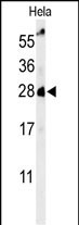
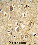
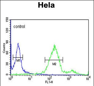
Application
| WB, IHC-P, FC, E |
|---|---|
| Primary Accession | P61006 |
| Reactivity | Human |
| Host | Rabbit |
| Clonality | Polyclonal |
| Isotype | Rabbit IgG |
| Calculated MW | 23668 Da |
| Gene ID | 4218 |
|---|---|
| Other Names | Ras-related protein Rab-8A, Oncogene c-mel, RAB8A, MEL, RAB8 |
| Target/Specificity | This RAB8A antibody is generated from rabbits immunized with human RAB8A recombinant protein. |
| Dilution | WB~~1:1000 IHC-P~~1:100~500 FC~~1:10~50 E~~Use at an assay dependent concentration. |
| Format | Purified polyclonal antibody supplied in PBS with 0.09% (W/V) sodium azide. This antibody is purified through a protein A column, followed by peptide affinity purification. |
| Storage | Maintain refrigerated at 2-8°C for up to 2 weeks. For long term storage store at -20°C in small aliquots to prevent freeze-thaw cycles. |
| Precautions | RAB8A Antibody is for research use only and not for use in diagnostic or therapeutic procedures. |
| Name | RAB8A (HGNC:7007) |
|---|---|
| Synonyms | MEL, RAB8 |
| Function | The small GTPases Rab are key regulators of intracellular membrane trafficking, from the formation of transport vesicles to their fusion with membranes. Rabs cycle between an inactive GDP-bound form and an active GTP-bound form that is able to recruit to membranes different sets of downstream effectors directly responsible for vesicle formation, movement, tethering and fusion. RAB8A is involved in polarized vesicular trafficking and neurotransmitter release. Together with RAB11A, RAB3IP, the exocyst complex, PARD3, PRKCI, ANXA2, CDC42 and DNMBP promotes transcytosis of PODXL to the apical membrane initiation sites (AMIS), apical surface formation and lumenogenesis (PubMed:20890297). Regulates the compacted morphology of the Golgi (PubMed:26209634). Together with MYO5B and RAB11A participates in epithelial cell polarization (PubMed:21282656). Also involved in membrane trafficking to the cilium and ciliogenesis (PubMed:21844891, PubMed:30398148, PubMed:20631154). Together with MICALL2, may also regulate adherens junction assembly (By similarity). May play a role in insulin-induced transport to the plasma membrane of the glucose transporter GLUT4 and therefore play a role in glucose homeostasis (By similarity). Involved in autophagy (PubMed:27103069). Participates in the export of a subset of neosynthesized proteins through a Rab8-Rab10- Rab11-dependent endososomal export route (PubMed:32344433). Targeted to and stabilized on stressed lysosomes through LRRK2 phosphorylation (PubMed:30209220). Suppresses stress-induced lysosomal enlargement through EHBP1 and EHNP1L1 effector proteins (PubMed:30209220). |
| Cellular Location | Cell membrane; Lipid-anchor; Cytoplasmic side. Golgi apparatus. Endosome membrane. Recycling endosome membrane. Cell projection, cilium. Cytoplasmic vesicle, phagosome. Cytoplasmic vesicle, phagosome membrane {ECO:0000250|UniProtKB:Q92930}; Lipid-anchor {ECO:0000250|UniProtKB:Q92930}; Cytoplasmic side {ECO:0000250|UniProtKB:Q92930}. Cytoplasm, cytoskeleton, microtubule organizing center, centrosome, centriole {ECO:0000250|UniProtKB:P55258}. Cytoplasm, cytoskeleton, cilium basal body. Midbody. Cytoplasm, cytoskeleton, cilium axoneme. Cytoplasm Lysosome. Note=Colocalizes with OPTN at the Golgi complex and in vesicular structures close to the plasma membrane (PubMed:15837803). In the GDP-bound form, present in the perinuclear region (PubMed:12221131). Shows a polarized distribution to distal regions of cell protrusions in the GTP-bound form (PubMed:12221131). Colocalizes with PARD3, PRKCI, EXOC5, OCLN, PODXL and RAB11A in apical membrane initiation sites (AMIS) during the generation of apical surface and lumenogenesis (PubMed:20890297) Localizes to tubular recycling endosome (PubMed:19864458). Recruited to phagosomes containing S.aureus or M.tuberculosis (PubMed:21255211) Non-phosphorylated RAB8A predominantly localized to the cytoplasm whereas phosphorylated RAB8A localized to the membrane (PubMed:26824392, PubMed:29125462, PubMed:30398148). Colocalized with MICAL1, GRAF1/ARHGAP26 and GRAF2/ARHGAP10 on endosomal tubules (PubMed:32344433). Localizes to enlarged lysosomes through LRRK2 phosphorylation (PubMed:30209220). Colocalizes with RPGR at the primary cilia of epithelial cells (By similarity) {ECO:0000250|UniProtKB:P61007, ECO:0000269|PubMed:12221131, ECO:0000269|PubMed:15837803, ECO:0000269|PubMed:19864458, ECO:0000269|PubMed:20890297, ECO:0000269|PubMed:21255211, ECO:0000269|PubMed:26824392, ECO:0000269|PubMed:29125462, ECO:0000269|PubMed:30209220, ECO:0000269|PubMed:30398148, ECO:0000269|PubMed:32344433} |
For Research Use Only. Not For Use In Diagnostic Procedures.

Provided below are standard protocols that you may find useful for product applications.
BACKGROUND
The proteasome is a multicatalytic proteinase complex with a highly ordered ring-shaped 20S core structure. The core structure is composed of 4 rings of 28 non-identical subunits; 2 rings are composed of 7 alpha subunits and 2 rings are composed of 7 beta subunits. Proteasomes are distributed throughout eukaryotic cells at a high concentration and cleave peptides in an ATP/ubiquitin-dependent process in a non-lysosomal pathway. Anessential function of a modified proteasome, the immunoproteasome, is the processing of class I MHC peptides. RAB8A is a member of the peptidase T1A family, that is a 20S core alpha subunit. A pseudogene has been identified on the Y chromosome.
REFERENCES
Kato,S., et.al., Gene 150 (2), 243-250 (1994)
Bey,F., et.al., Mol. Gen. Genet. 237 (1-2), 193-205 (1993)
终于等到您。ABCEPTA(百远生物)抗体产品。
点击下方“我要评价 ”按钮提交您的反馈信息,您的反馈和评价是我们最宝贵的财富之一,
我们将在1-3个工作日内处理您的反馈信息。
如有疑问,联系:0512-88856768 tech-china@abcepta.com.


