


 癌症的基本特征包括细胞增殖、血管生成、迁移、凋亡逃避机制和细胞永生等。找到癌症发生过程中这些通路的关键标记物和对应的抗体用于检测至关重要。
 为您推荐一个泛素化位点预测神器——泛素化分析工具,可以为您的蛋白的泛素化位点作出预测和评分。
 细胞自噬受体图形绘图工具为你的蛋白的细胞受体结合位点作出预测和评分,识别结合到自噬通路中的蛋白是非常重要的,便于让我们理解自噬在正常生理、病理过程中的作用,如发育、细胞分化、神经退化性疾病、压力条件下、感染和癌症。
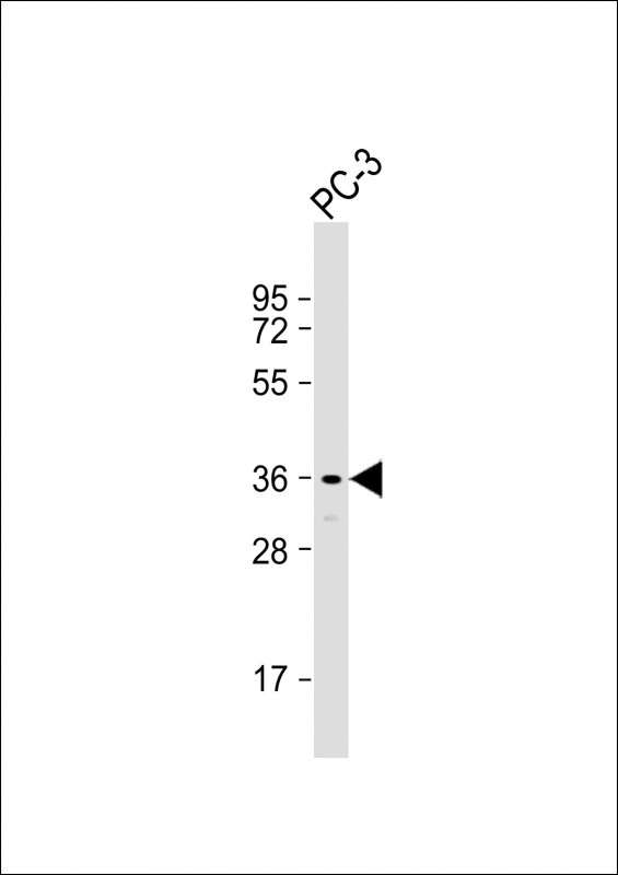
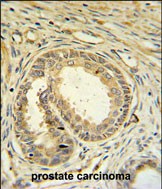
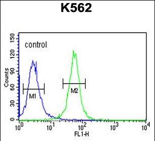
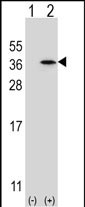
SPDYA Antibody (Center)
Affinity Purified Rabbit Polyclonal Antibody (Pab)
- 产品详情
- 文献引用 : 1
- 实验流程
- 背景知识
Application
| IHC-P, FC, WB, E |
|---|---|
| Primary Accession | Q5MJ70 |
| Other Accession | Q8R496, Q5IBH7 |
| Reactivity | Human |
| Predicted | Mouse, Rat |
| Host | Rabbit |
| Clonality | Polyclonal |
| Isotype | Rabbit IgG |
| Calculated MW | 36463 Da |
| Antigen Region | 147-175 aa |
| Gene ID | 245711 |
|---|---|
| Other Names | Speedy protein A, Rapid inducer of G2/M progression in oocytes A, RINGO A, hSpy/Ringo A, Speedy-1, Spy1, SPDYA (HGNC:30613) |
| Target/Specificity | This SPDYA antibody is generated from rabbits immunized with a KLH conjugated synthetic peptide between 147-175 amino acids from the Central region of human SPDYA. |
| Dilution | IHC-P~~1:100~500 FC~~1:10~50 WB~~1:1000 E~~Use at an assay dependent concentration. |
| Format | Purified polyclonal antibody supplied in PBS with 0.09% (W/V) sodium azide. This antibody is purified through a protein A column, followed by peptide affinity purification. |
| Storage | Maintain refrigerated at 2-8°C for up to 2 weeks. For long term storage store at -20°C in small aliquots to prevent freeze-thaw cycles. |
| Precautions | SPDYA Antibody (Center) is for research use only and not for use in diagnostic or therapeutic procedures. |
| Name | SPDYA (HGNC:30613) |
|---|---|
| Function | Regulates the G1/S phase transition of the cell cycle by binding and activating CDK1 and CDK2 (PubMed:12972555). Contributes to CDK2 activation without promoting CDK2 phosphorylation, by inducing a conformation change of the CDK2 T-loop that obstructs the substrate- binding cleft prior to kinase activation (PubMed:28666995). Mediates cell survival during the DNA damage process through activation of CDK2 (PubMed:12839962). |
| Cellular Location | Nucleus |
| Tissue Location | Highly expressed in testis. Expressed at a low level in wide range of tissues including bone marrow, brain, heart, kidney, colon, liver, placenta, spleen, skeletal muscle, salivary gland, thyroid gland, thymus, trachea and uterus. Expressed at a slightly higher level in adrenal gland, cerebellum, small intestine, lung, prostate and trachea. Expression is cell cycle-dependent, being restricted to cells in G1/S phase. |
For Research Use Only. Not For Use In Diagnostic Procedures.

Provided below are standard protocols that you may find useful for product applications.
BACKGROUND
SPDYA (speedy homolog A (Drosophila)) regulates the G1/S phase transition of the cell cycle by binding and activating CDC2, CDK2 and CDKN1B/KIP1. SPDYA can activate CDK2 without promoting CDK2 phosphorylation. SPDYA mediates cell survival during the DNA damage process through activation of CDK2.
REFERENCES
Ke, Q., et al. Exp. Mol. Pathol. 87(3):167-172(2009)
Dinarina, A., et al. FEBS Lett. 583(17):2772-2778(2009)
McAndrew, C.W., et al. Cell Cycle 8(1):66-75(2009)
终于等到您。ABCEPTA(百远生物)抗体产品。
点击下方“我要评价 ”按钮提交您的反馈信息,您的反馈和评价是我们最宝贵的财富之一,
我们将在1-3个工作日内处理您的反馈信息。
如有疑问,联系:0512-88856768 tech-china@abcepta.com.


