


 癌症的基本特征包括细胞增殖、血管生成、迁移、凋亡逃避机制和细胞永生等。找到癌症发生过程中这些通路的关键标记物和对应的抗体用于检测至关重要。
 为您推荐一个泛素化位点预测神器——泛素化分析工具,可以为您的蛋白的泛素化位点作出预测和评分。
 细胞自噬受体图形绘图工具为你的蛋白的细胞受体结合位点作出预测和评分,识别结合到自噬通路中的蛋白是非常重要的,便于让我们理解自噬在正常生理、病理过程中的作用,如发育、细胞分化、神经退化性疾病、压力条件下、感染和癌症。
Anti-Caspase 8 Picoband Antibody
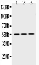
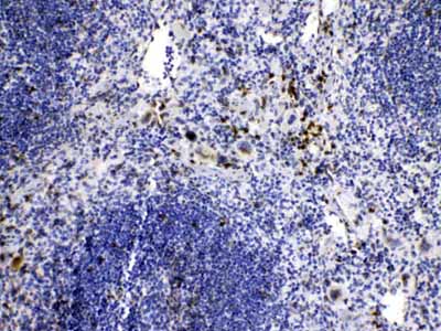
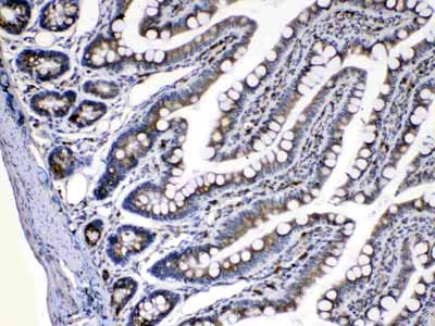
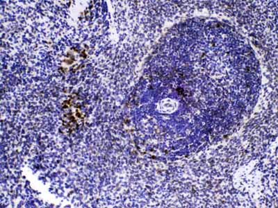
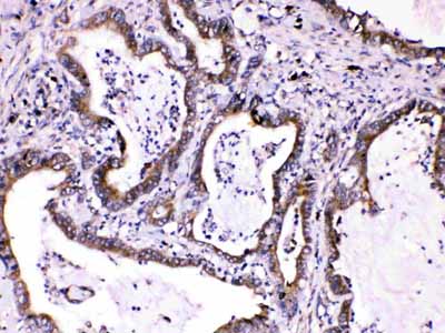
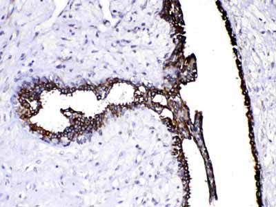
- 产品详情
- 实验流程
- 背景知识
Application 
| WB, IHC-P |
|---|---|
| Primary Accession | Q14790 |
| Host | Rabbit |
| Reactivity | Human, Mouse, Rat |
| Clonality | Polyclonal |
| Format | Lyophilized |
| Description | Rabbit IgG polyclonal antibody for Caspase-8(CASP8) detection. Tested with WB, IHC-P in Human;Mouse;Rat. |
| Reconstitution | Add 0.2ml of distilled water will yield a concentration of 500ug/ml. |
| Gene ID | 841 |
|---|---|
| Other Names | Caspase-8, CASP-8, 3.4.22.61, Apoptotic cysteine protease, Apoptotic protease Mch-5, CAP4, FADD-homologous ICE/ced-3-like protease, FADD-like ICE, FLICE, ICE-like apoptotic protease 5, MORT1-associated ced-3 homolog, MACH, Caspase-8 subunit p18, Caspase-8 subunit p10, CASP8, MCH5 |
| Calculated MW | 55391 Da |
| Application Details | Immunohistochemistry(Paraffin-embedded Section), 0.5-1 µg/ml, Human, Mouse, Rat, By Heat Western blot, 0.1-0.5 µg/ml, Human, Mouse, Rat |
| Subcellular Localization | Cytoplasm. |
| Tissue Specificity | Isoform 1, isoform 5 and isoform 7 are expressed in a wide variety of tissues. Highest expression in peripheral blood leukocytes, spleen, thymus and liver. Barely detectable in brain, testis and skeletal muscle. |
| Source | Eukaryota |
| Protein Name | Caspase-8 |
| Contents | Each vial contains 5mg BSA, 0.9mg NaCl, 0.2mg Na2HPO4, 0.05mg NaN3. |
| Immunogen | A synthetic peptide corresponding to a sequence at the N-terminus of human Caspase 8 (410-449aa VSYRNPAEGTWYIQSLCQSLRERCPRGDDILTILTEVNYE), different from the related mouse and rat sequences by seven amino acids. |
| Purification | Immunogen affinity purified. |
| Cross Reactivity | No cross reactivity with other proteins |
| Storage | At -20˚C for one year. After r˚Constitution, at 4˚C for one month. It˚Can also be aliquotted and stored frozen at -20˚C for a longer time.Avoid repeated freezing and thawing. |
For Research Use Only. Not For Use In Diagnostic Procedures.
| Name | CASP8 {ECO:0000303|PubMed:9931493, ECO:0000312|HGNC:HGNC:1509} |
|---|---|
| Function | Thiol protease that plays a key role in programmed cell death by acting as a molecular switch for apoptosis, necroptosis and pyroptosis, and is required to prevent tissue damage during embryonic development and adulthood (PubMed:23516580, PubMed:35338844, PubMed:35446120, PubMed:8681376, PubMed:8681377, PubMed:8962078, PubMed:9006941, PubMed:9184224). Initiator protease that induces extrinsic apoptosis by mediating cleavage and activation of effector caspases responsible for FAS/CD95-mediated and TNFRSF1A-induced cell death (PubMed:23516580, PubMed:35338844, PubMed:35446120, PubMed:8681376, PubMed:8681377, PubMed:8962078, PubMed:9006941, PubMed:9184224). Cleaves and activates effector caspases CASP3, CASP4, CASP6, CASP7, CASP9 and CASP10 (PubMed:16916640, PubMed:8962078, PubMed:9006941). Binding to the adapter molecule FADD recruits it to either receptor FAS/TNFRSF6 or TNFRSF1A (PubMed:8681376, PubMed:8681377). The resulting aggregate called the death-inducing signaling complex (DISC) performs CASP8 proteolytic activation (PubMed:9184224). The active dimeric enzyme is then liberated from the DISC and free to activate downstream apoptotic proteases (PubMed:9184224). Proteolytic fragments of the N-terminal propeptide (termed CAP3, CAP5 and CAP6) are likely retained in the DISC (PubMed:9184224). Also cleaves and activates BID, thereby promoting cytochrome C release from mitochrondria (By similarity). In addition to extrinsic apoptosis, also acts as a negative regulator of necroptosis: acts by cleaving RIPK1 at 'Asp-324', which is crucial to inhibit RIPK1 kinase activity, limiting TNF-induced apoptosis, necroptosis and inflammatory response (PubMed:31827280, PubMed:31827281). Also able to initiate pyroptosis by mediating cleavage and activation of gasdermin-C and -D (GSDMC and GSDMD, respectively): gasdermin cleavage promotes release of the N-terminal moiety that binds to membranes and forms pores, triggering pyroptosis (PubMed:32929201, PubMed:34012073). Initiates pyroptosis following inactivation of MAP3K7/TAK1 (By similarity). Also acts as a regulator of innate immunity by mediating cleavage and inactivation of N4BP1 downstream of TLR3 or TLR4, thereby promoting cytokine production (By similarity). May participate in the Granzyme B (GZMB) cell death pathways (PubMed:8755496). Cleaves PARP1 and PARP2 (PubMed:8681376). Independent of its protease activity, promotes cell migration following phosphorylation at Tyr-380 (PubMed:18216014, PubMed:27109099). |
| Cellular Location | Cytoplasm {ECO:0000250|UniProtKB:Q9JHX4}. Nucleus {ECO:0000250|UniProtKB:Q9JHX4}. Cell projection, lamellipodium. Note=Recruitment to lamellipodia of migrating cells is enhanced by phosphorylation at Tyr-380 |
| Tissue Location | Isoform 1, isoform 5 and isoform 7 are expressed in a wide variety of tissues. Highest expression in peripheral blood leukocytes, spleen, thymus and liver. Barely detectable in brain, testis and skeletal muscle |
Provided below are standard protocols that you may find useful for product applications.
BACKGROUND
CASP8 is also known as CAP4, MACH or MCH5. This gene encodes a member of the cysteine-aspartic acid protease (caspase) family. Sequential activation of caspases plays a central role in the execution-phase of cell apoptosis. Caspases exist as inactive proenzymes composed of a prodomain, a large protease subunit, and a small protease subunit. Activation of caspases requires proteolytic processing at conserved internal aspartic residues to generate a heterodimeric enzyme consisting of the large and small subunits. This protein is involved in the programmed cell death induced by Fas and various apoptotic stimuli. The N-terminal FADD-like death effector domain of this protein suggests that it may interact with Fas-interacting protein FADD. In addtion, this protein was detected in the insoluble fraction of the affected brain region from Huntington disease patients but not in those from normal controls, which implicated the role in neurodegenerative diseases. Many alternatively spliced transcript variants encoding different isoforms have been described, although not all variants have had their full-length sequences determined.

终于等到您。ABCEPTA(百远生物)抗体产品。
点击下方“我要评价 ”按钮提交您的反馈信息,您的反馈和评价是我们最宝贵的财富之一,
我们将在1-3个工作日内处理您的反馈信息。
如有疑问,联系:0512-88856768 tech-china@abcepta.com.


