


 癌症的基本特征包括细胞增殖、血管生成、迁移、凋亡逃避机制和细胞永生等。找到癌症发生过程中这些通路的关键标记物和对应的抗体用于检测至关重要。
 为您推荐一个泛素化位点预测神器——泛素化分析工具,可以为您的蛋白的泛素化位点作出预测和评分。
 细胞自噬受体图形绘图工具为你的蛋白的细胞受体结合位点作出预测和评分,识别结合到自噬通路中的蛋白是非常重要的,便于让我们理解自噬在正常生理、病理过程中的作用,如发育、细胞分化、神经退化性疾病、压力条件下、感染和癌症。
Anti-P53 Antibody
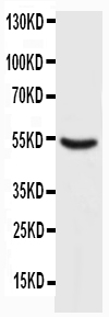
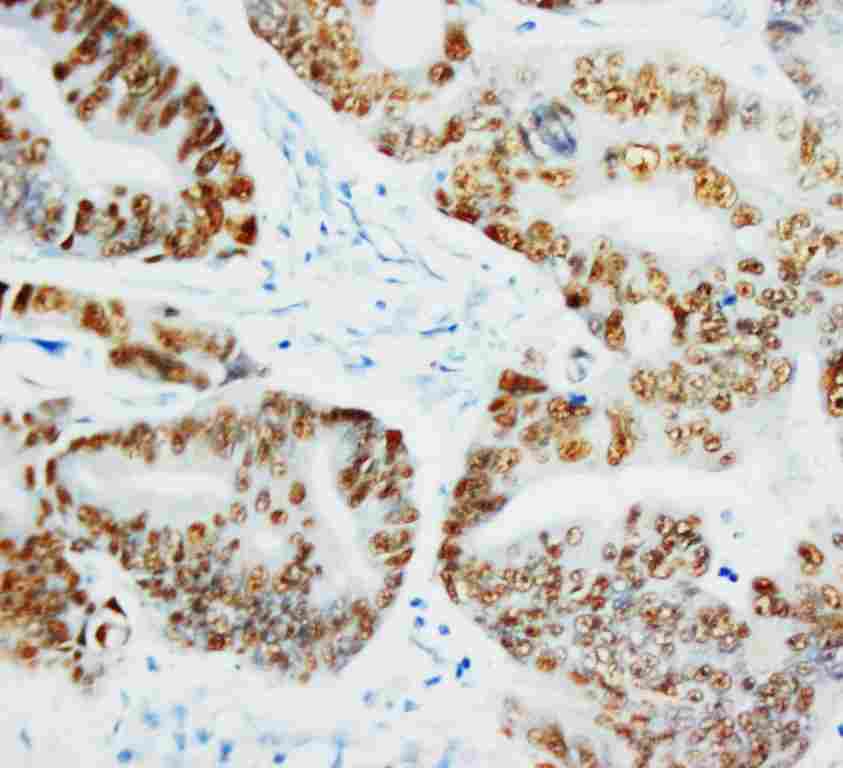
- 产品详情
- 实验流程
- 背景知识
Application 
| WB, IHC-P, IHC-F, ICC |
|---|---|
| Primary Accession | P10361 |
| Host | Mouse |
| Isotype | Mouse IgG2a |
| Reactivity | Human |
| Clonality | Monoclonal |
| Format | Lyophilized |
| Description | Mouse IgG monoclonal antibody for p53, tumor protein p53 (TP53) detection. Tested with WB, IHC-P, IHC-F, ICC in Human. No cross reactivity with other proteins. |
| Reconstitution | Add 1ml of PBS buffer will yield a concentration of 100ug/ml. |
| Gene ID | 24842 |
|---|---|
| Other Names | Cellular tumor antigen p53, Tumor suppressor p53, Tp53, P53 |
| Calculated MW | 43451 Da |
| Application Details | Immunohistochemistry(Paraffin-embedded Section), 0.5-2 µg/ml, Human, By Heat Immunocytochemistry , 1 µg/ml, Human, - Immunohistochemistry(Frozen Section), 0.5-2 µg/ml, Human, - Western blot, 0.5-1 µg/ml, Human |
| Subcellular Localization | Cytoplasm . Nucleus . Endoplasmic reticulum . Mitochondrion matrix . Interaction with BANP promotes nuclear localization. Translocates to mitochondria upon oxidative stress (By similarity). . |
| Tissue Specificity | TP53: Ubiquitous. Isoforms are expressed in a widerange of normal tissues but in a tissue-dependent manner. Isoform2 is expressed in most normal tissues but is not detected inbrain, lung, prostate, muscle, fetal brain, spinal cord and fetalliver. Isoform 3 is expressed in most normal tissues but is notdetected in lung, spleen, testis, fetal brain, spinal cord andfetal liver. Isoform 7 is expressed in most normal tissues but isnot detected in prostate, uterus, skeletal muscle and breast.Isoform 8 is detected only in colon, bone marrow, testis, fetalbrain and intestine. Isoform 9 is expressed in most normal tissuesbut is not detected in brain, heart, lung, fetal liver, salivarygland, breast or intestine. |
| Source | Eukaryota |
| Protein Name | Cellular tumor antigen p53 |
| Contents | Mouse ascites fluid, 1.2% sodium acetate, 2mg BSA, with 0.01mg NaN3 as preservative. |
| Clone Names | BP53-12 |
| Immunogen | Recombinant human wild-type p53 protein. |
| Purification | Ascites |
| Cross Reactivity | No cross reactivity with other proteins |
| Storage | At -20˚C for one year. After r˚Constitution, at 4˚C for one month. It˚Can also be aliquotted and stored frozen at -20˚C for a longer time.Avoid repeated freezing and thawing. |
| Sequence Similarities | Belongs to the p53 family. |
For Research Use Only. Not For Use In Diagnostic Procedures.
| Name | Tp53 |
|---|---|
| Synonyms | P53 |
| Function | Multifunctional transcription factor that induces cell cycle arrest, DNA repair or apoptosis upon binding to its target DNA sequence. Acts as a tumor suppressor in many tumor types; induces growth arrest or apoptosis depending on the physiological circumstances and cell type. Negatively regulates cell division by controlling expression of a set of genes required for this process. One of the activated genes is an inhibitor of cyclin-dependent kinases. Apoptosis induction seems to be mediated either by stimulation of BAX and FAS antigen expression, or by repression of Bcl-2 expression. Its pro- apoptotic activity is activated via its interaction with PPP1R13B/ASPP1 or TP53BP2/ASPP2 (By similarity). However, this activity is inhibited when the interaction with PPP1R13B/ASPP1 or TP53BP2/ASPP2 is displaced by PPP1R13L/iASPP (By similarity). In cooperation with mitochondrial PPIF is involved in activating oxidative stress-induced necrosis; the function is largely independent of transcription. Prevents CDK7 kinase activity when associated to CAK complex in response to DNA damage, thus stopping cell cycle progression. Induces the transcription of long intergenic non-coding RNA p21 (lincRNA-p21) and lincRNA-Mkln1. LincRNA- p21 participates in TP53-dependent transcriptional repression leading to apoptosis and seems to have an effect on cell-cycle regulation. Regulates the circadian clock by repressing CLOCK-ARNTL/BMAL1-mediated transcriptional activation of PER2. |
| Cellular Location | Cytoplasm {ECO:0000250|UniProtKB:P04637}. Nucleus {ECO:0000250|UniProtKB:P04637}. Nucleus, PML body {ECO:0000250|UniProtKB:P04637}. Endoplasmic reticulum {ECO:0000250|UniProtKB:P04637}. Mitochondrion matrix {ECO:0000250|UniProtKB:P04637}. Cytoplasm, cytoskeleton, microtubule organizing center, centrosome {ECO:0000250|UniProtKB:P04637} Note=Interaction with BANP promotes nuclear localization. Recruited into PML bodies together with CHEK2. Translocates to mitochondria upon oxidative stress. Translocates to mitochondria in response to mitomycin C treatment (By similarity). Competitive inhibition of TP53 interaction with HSPA9/MOT-2 by UBXN2A results in increased protein abundance and subsequent translocation of TP53 to the nucleus (By similarity) {ECO:0000250|UniProtKB:P04637} |
Provided below are standard protocols that you may find useful for product applications.
BACKGROUND
The p53 tumor antigen is found in increased amounts in a wide variety of transformed cells. The protein is also detectable in many actively proliferating, nontransformed cells, but it is undetectable or present at low levels in resting cells. This protein induces cell cycle arrest or apoptosis in response to sublethal or severe DNA damage, respectively, by differential transcription of target genes and through transcription-independent apoptotic functions. The p53 protein contains 393 amino acids. Human p53 tumour antigen is Locatedto band 17p13. p53 mutations are common in pancreatic cancer and are absent in chronic pancreatitis.

终于等到您。ABCEPTA(百远生物)抗体产品。
点击下方“我要评价 ”按钮提交您的反馈信息,您的反馈和评价是我们最宝贵的财富之一,
我们将在1-3个工作日内处理您的反馈信息。
如有疑问,联系:0512-88856768 tech-china@abcepta.com.


