


 癌症的基本特征包括细胞增殖、血管生成、迁移、凋亡逃避机制和细胞永生等。找到癌症发生过程中这些通路的关键标记物和对应的抗体用于检测至关重要。
 为您推荐一个泛素化位点预测神器——泛素化分析工具,可以为您的蛋白的泛素化位点作出预测和评分。
 细胞自噬受体图形绘图工具为你的蛋白的细胞受体结合位点作出预测和评分,识别结合到自噬通路中的蛋白是非常重要的,便于让我们理解自噬在正常生理、病理过程中的作用,如发育、细胞分化、神经退化性疾病、压力条件下、感染和癌症。
Anti-GBP1 Antibody
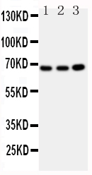
- 产品详情
- 实验流程
- 背景知识
Application 
| WB |
|---|---|
| Primary Accession | P32455 |
| Host | Rabbit |
| Reactivity | Human |
| Clonality | Polyclonal |
| Format | Lyophilized |
| Description | Rabbit IgG polyclonal antibody for Guanylate-binding protein 1(GBP1) detection. Tested with WB in Human. |
| Reconstitution | Add 0.2ml of distilled water will yield a concentration of 500ug/ml. |
| Gene ID | 2633 |
|---|---|
| Other Names | Guanylate-binding protein 1, 3.6.5.-, GTP-binding protein 1, GBP-1, HuGBP-1, Guanine nucleotide-binding protein 1, Interferon-induced guanylate-binding protein 1, GBP1 |
| Calculated MW | 67931 Da |
| Application Details | Western blot, 0.1-0.5 µg/ml, Human |
| Subcellular Localization | Cytoplasm. Golgi apparatus membrane; Lipid- anchor; Cytoplasmic side. Secreted. Secreted from endothelial cells in the cerebrospinal fluid, upon bacterial challenge and independently of interferon-gamma induction. Golgi membrane localization requires isoprenylation and the presence of another IFN-gamma-induced factor. |
| Source | Eukaryota |
| Protein Name | Guanylate-binding protein 1 |
| Contents | Each vial contains 5mg BSA, 0.9mg NaCl, 0.2mg Na2HPO4, 0.05mg Thimerosal, 0.05mg NaN3. |
| Immunogen | A synthetic peptide corresponding to a sequence at the C-terminus of human GBP1(566-589aa QKESRIMKNEIQDLQTKMRRRKAC). |
| Purification | Immunogen affinity purified. |
| Cross Reactivity | No cross reactivity with other proteins |
| Storage | At -20˚C for one year. After r˚Constitution, at 4˚C for one month. It˚Can also be aliquotted and stored frozen at -20˚C for a longer time.Avoid repeated freezing and thawing. |
| Sequence Similarities | Belongs to the TRAFAC class dynamin-like GTPase superfamily. GB1/RHD3-type GTPase family. GB1 subfamily. |
For Research Use Only. Not For Use In Diagnostic Procedures.
| Name | GBP1 {ECO:0000303|PubMed:7512561, ECO:0000312|HGNC:HGNC:4182} |
|---|---|
| Function | Interferon (IFN)-inducible GTPase that plays important roles in innate immunity against a diverse range of bacterial, viral and protozoan pathogens (PubMed:16511497, PubMed:22106366, PubMed:29144452, PubMed:31268602, PubMed:32510692, PubMed:32581219, PubMed:37797010, PubMed:7512561). Hydrolyzes GTP to GMP in two consecutive cleavage reactions: GTP is first hydrolyzed to GDP and then to GMP in a processive manner (PubMed:16511497, PubMed:32510692, PubMed:7512561, PubMed:39394410). Following infection, recruited to the pathogen- containing vacuoles or vacuole-escaped bacteria and promotes both inflammasome assembly and autophagy (PubMed:29144452, PubMed:31268602). Acts as a positive regulator of inflammasome assembly by facilitating the detection of inflammasome ligands from pathogens (PubMed:31268602, PubMed:32510692, PubMed:32581219). Involved in the lysis of pathogen- containing vacuoles, releasing pathogens into the cytosol (By similarity). Following pathogen release in the cytosol, forms a protein coat in a GTPase-dependent manner that encapsulates pathogens and promotes the detection of ligands by pattern recognition receptors (PubMed:32510692, PubMed:32581219). Plays a key role in inflammasome assembly in response to infection by Gram-negative bacteria: following pathogen release in the cytosol, forms a protein coat that encapsulates Gram-negative bacteria and directly binds to lipopolysaccharide (LPS), disrupting the O-antigen barrier and unmasking lipid A that is that detected by the non-canonical inflammasome effector CASP4/CASP11 (PubMed:32510692, PubMed:32581219). Also promotes recruitment of proteins that mediate bacterial cytolysis, leading to release double- stranded DNA (dsDNA) that activates the AIM2 inflammasome (PubMed:31268602). Involved in autophagy by regulating bacteriolytic peptide generation via its interaction with ubiquitin-binding protein SQSTM1, which delivers monoubiquitinated proteins to autolysosomes for the generation of bacteriolytic peptides (By similarity). Confers protection to several pathogens, including the bacterial pathogens L.monocytogenes and M.bovis BCG as well as the protozoan pathogen T.gondii (PubMed:31268602). Exhibits antiviral activity against influenza virus (PubMed:22106366). |
| Cellular Location | Cytoplasmic vesicle membrane; Lipid-anchor; Cytoplasmic side. Golgi apparatus membrane; Lipid-anchor; Cytoplasmic side. Cell membrane; Lipid-anchor; Cytoplasmic side. Cytoplasm, cytosol. Secreted. Note=Localizes to pathogen-containing vacuoles or to the cell surface of bacteria that escaped vacuoles (PubMed:29144452, PubMed:31268602, PubMed:32510692, PubMed:32581219) Secreted from endothelial cells in the cerebrospinal fluid, upon bacterial challenge and independently of IFNG induction (PubMed:16936281). Golgi membrane localization requires isoprenylation and the presence of another IFNG-induced factor (PubMed:15937107) Sequestered in the cytosol following phosphorylation by PIM1 and subsequent interaction with 14-3-3 protein sigma (SFN) (PubMed:37797010). |
Provided below are standard protocols that you may find useful for product applications.
BACKGROUND
Interferons induce a large number of genes in their target cells, including those coding for guanylate-binding proteins(GBPs). GBPs, such as GBP1, are characterized by their ability to specifically bind guanine nucleotides(GMP, GDP, and GTP) and are distinguished from the GTP-binding proteins by the presence of 2 binding motifs rather than 3. The 593-amino acid GBP1 protein shares 77% and 88% identity with GBP2 and GBP3, respectively. All GBPs, including GBP1, have a conserved N-terminal globular GTP-binding domain containing 2 consensus sequences and a third T(L/V)RD sequence not found in other GTPases. The GBP1 gene to the GBP gene cluster on chromosome 1p22.2. It is located telomeric to GBP2 and centromeric to GBP3. High expression of GBP1, GBP2, and GBP3 are in endothelial cells after stimulation with IFNG, TNF, or IL1B .

终于等到您。ABCEPTA(百远生物)抗体产品。
点击下方“我要评价 ”按钮提交您的反馈信息,您的反馈和评价是我们最宝贵的财富之一,
我们将在1-3个工作日内处理您的反馈信息。
如有疑问,联系:0512-88856768 tech-china@abcepta.com.


