


 癌症的基本特征包括细胞增殖、血管生成、迁移、凋亡逃避机制和细胞永生等。找到癌症发生过程中这些通路的关键标记物和对应的抗体用于检测至关重要。
 为您推荐一个泛素化位点预测神器——泛素化分析工具,可以为您的蛋白的泛素化位点作出预测和评分。
 细胞自噬受体图形绘图工具为你的蛋白的细胞受体结合位点作出预测和评分,识别结合到自噬通路中的蛋白是非常重要的,便于让我们理解自噬在正常生理、病理过程中的作用,如发育、细胞分化、神经退化性疾病、压力条件下、感染和癌症。
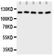
Anti-Desmoglein 2 Antibody
- 产品详情
- 实验流程
- 背景知识
Application 
| WB, IHC-P |
|---|---|
| Primary Accession | Q14126 |
| Host | Rabbit |
| Reactivity | Human, Mouse, Rat |
| Clonality | Polyclonal |
| Format | Lyophilized |
| Description | Rabbit IgG polyclonal antibody for Desmoglein-2(DSG2) detection. Tested with WB, IHC-P in Human;Mouse;Rat. |
| Reconstitution | Add 0.2ml of distilled water will yield a concentration of 500ug/ml. |
| Gene ID | 1829 |
|---|---|
| Other Names | Desmoglein-2, Cadherin family member 5, HDGC, DSG2, CDHF5 |
| Calculated MW | 122294 Da |
| Application Details | Immunohistochemistry(Paraffin-embedded Section), 0.5-1 µg/ml, Human, Rat, Mouse, By Heat Western blot, 0.1-0.5 µg/ml, Human, Rat, Mouse |
| Subcellular Localization | Cell membrane; Single-pass type I membrane protein. Cell junction, desmosome. |
| Tissue Specificity | All of the tissues tested and carcinomas. |
| Source | Eukaryota |
| Protein Name | Desmoglein-2 |
| Contents | Each vial contains 5mg BSA, 0.9mg NaCl, 0.2mg Na2HPO4, 0.05mg Thimerosal, 0.05mg NaN3. |
| Immunogen | A synthetic peptide corresponding to a sequence at the C-terminus of human Desmoglein 2(1104-1118aa STRVTKHSTVQHSYS), different from the related mouse and rat sequences by one amino acid. |
| Purification | Immunogen affinity purified. |
| Cross Reactivity | No cross reactivity with other proteins |
| Storage | At -20˚C for one year. After r˚Constitution, at 4˚C for one month. It˚Can also be aliquotted and stored frozen at -20˚C for a longer time.Avoid repeated freezing and thawing. |
| Sequence Similarities | Contains 4 cadherin domains. |
For Research Use Only. Not For Use In Diagnostic Procedures.
| Name | DSG2 |
|---|---|
| Synonyms | CDHF5 |
| Function | A component of desmosome cell-cell junctions which are required for positive regulation of cellular adhesion (PubMed:17559062, PubMed:38395410). Involved in the interaction of plaque proteins and intermediate filaments mediating cell-cell adhesion. Required for proliferation and viability of embryonic stem cells in the blastocyst, thereby crucial for progression of post-implantation embryonic development (By similarity). Maintains pluripotency by regulating epithelial to mesenchymal transition/mesenchymal to epithelial transition (EMT/MET) via interacting with and sequestering CTNNB1 to sites of cell-cell contact, thereby reducing translocation of CTNNB1 to the nucleus and subsequent transcription of CTNNB1/TCF-target genes (PubMed:29910125). Promotes pluripotency and the multi-lineage differentiation potential of hematopoietic stem cells (PubMed:27338829). Plays a role in endothelial cell sprouting and elongation via mediating the junctional-association of cortical actin fibers and CDH5 (PubMed:27338829). Promotes cardiomyocyte cell homeostasis and desmosome junction formation at intercalated disks, as a result plays a role in the maintenance of cardiac conduction and heart chamber integrity (By similarity). Positively regulates pancreatic islet development and maintenance of endothelial cell barrier integrity in the pancreas, therefore involved in the controlled release of insulin from islet cells into the circulation in response to glucose (By similarity). Plays a role in limiting inflammatory infiltration and the apoptotic response to injury in kidney tubular epithelial cells, potentially via its role in maintaining cell-cell adhesion and the epithelial barrier (PubMed:38395410). Acts as a positive modulator of CSK and EGFR activation via sequestering them away from lipid rafts, this is independent of its role in desmosome cell junctions (PubMed:26918609). Also disrupts the localization of CAV1 to lipid rafts resulting in its distribution throughout the cytoplasm (PubMed:26918609). |
| Cellular Location | Cell membrane; Single-pass type I membrane protein. Cell junction, desmosome. Cytoplasm. Note=Localized to intercalated disks in the heart (PubMed:31845994). Localizes to the cytoplasm following cleavage by CASP3 in response to apoptosis (PubMed:17559062) Glycosylation promotes localization to the plasma membrane (PubMed:30885746). |
| Tissue Location | Expressed in undifferentiated pluripotent stem cells, expression decreases during differentiation (at protein level) (PubMed:29910125). Expressed in hematopoietic stem cells and circulating endothelial progenitor cells, expression decreases upon increasing cell lineage commitment (at protein level) (PubMed:27338829). Expressed on common myeloid progenitors, pro- myelocytes, pro-erythrocytes and B-cell linage progenitors (at protein level). Expression in mature cell types in the bone marrow and mature leukocyte populations is absent (PubMed:27338829). Expressed by foreskin fibroblasts, expression peaks during the early stage of differentiation reprogramming (at protein level) (PubMed:29910125) Expressed by endothelial cells in both arterioles and venules in the cervix (at protein level) (PubMed:27338829). Expressed in pancreatic alpha-cells, beta-cells and exocrine tissue (at protein level) (PubMed:36309486). Expressed in cardiomyocytes (at protein level) (PubMed:31845994, PubMed:38375917). Expressed in kidney tubular epithelial cells (PubMed:38395410). Expressed in the esophageal mucosa (PubMed:24220297). |
Provided below are standard protocols that you may find useful for product applications.
BACKGROUND
Desmoglein-2 is a protein that in humans is encoded by the DSG2 gene. These desmoglein gene family members are located in a cluster on chromosome 18. This second family member is expressed in colon, colon carcinoma, and other simple and stratified epithelial-derived cell lines. Mutations in DSG2 display a high degree of penetrance. Disease expression was of variable severity with LV involvement a prominent feature. The low prevalence of classical ECG changes highlights the need to expand current diagnostic criteria to take account of LV disease, childhood disease expression, and incomplete penetrance.

终于等到您。ABCEPTA(百远生物)抗体产品。
点击下方“我要评价 ”按钮提交您的反馈信息,您的反馈和评价是我们最宝贵的财富之一,
我们将在1-3个工作日内处理您的反馈信息。
如有疑问,联系:0512-88856768 tech-china@abcepta.com.


