


 癌症的基本特征包括细胞增殖、血管生成、迁移、凋亡逃避机制和细胞永生等。找到癌症发生过程中这些通路的关键标记物和对应的抗体用于检测至关重要。
 为您推荐一个泛素化位点预测神器——泛素化分析工具,可以为您的蛋白的泛素化位点作出预测和评分。
 细胞自噬受体图形绘图工具为你的蛋白的细胞受体结合位点作出预测和评分,识别结合到自噬通路中的蛋白是非常重要的,便于让我们理解自噬在正常生理、病理过程中的作用,如发育、细胞分化、神经退化性疾病、压力条件下、感染和癌症。
Anti-Aurora A Antibody
- 产品详情
- 实验流程
- 背景知识
Application 
| WB |
|---|---|
| Primary Accession | P97477 |
| Host | Rabbit |
| Reactivity | Mouse, Rat |
| Clonality | Polyclonal |
| Format | Lyophilized |
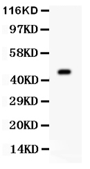
| Description | Rabbit IgG polyclonal antibody for Aurora kinase A(AURKA) detection. Tested with WB in Mouse;Rat. |
| Reconstitution | Add 0.2ml of distilled water will yield a concentration of 500ug/ml. |
| Gene ID | 20878 |
|---|---|
| Other Names | Aurora kinase A, 2.7.11.1, Aurora 2, Aurora family kinase 1, Aurora/IPL1-related kinase 1, ARK-1, Aurora-related kinase 1, Ipl1- and aurora-related kinase 1, Serine/threonine-protein kinase 6, Serine/threonine-protein kinase Ayk1, Serine/threonine-protein kinase aurora-A, Aurka |
| Calculated MW | 44772 Da |
| Application Details | Western blot, 0.1-0.5 µg/ml, Mouse, Rat |
| Subcellular Localization | Cytoplasm, cytoskeleton, microtubule organizing center, centrosome. Cytoplasm, cytoskeleton, spindle pole. Localizes on centrosomes in interphase cells and at each spindle pole in mitosis. Associates with both the pericentriolar material (PCM) and centrioles. Colocalized with SIRT2 at centrosome (By similarity). Detected at the neurite hillock in developing neurons. . |
| Tissue Specificity | Detected in embryonic neurons in dorsal root ganglia and brain cortex (at protein level). Highly expressed in testis, in about one third of the seminiferous tubules. Expression is restricted to specific spermatocytes nearing completion of prophase, with levels falling off on transition to elongated spermatids. Highly expressed in the ovary, expression in the oocyte starts around the transition to large growing follicle. Abundant expression is seen in the proliferating granulosa and thecal cells of the growing follicle, and in the young corpus luteum. Very weakly expressed in spleen and intestine. . |
| Source | Eukaryota |
| Protein Name | Aurora kinase A |
| Contents | Each vial contains 5mg BSA, 0.9mg NaCl, 0.2mg Na2HPO4, 0.05mg Thimerosal, 0.05mg NaN3. |
| Immunogen | A synthetic peptide corresponding to a sequence in the middle region of mouse Aurora A(109-125aa QKTEDTKKRQWTLEDFD), different from the related rat sequence by one amino acid. |
| Purification | Immunogen affinity purified. |
| Cross Reactivity | No cross reactivity with otherproteins |
| Storage | At -20˚C for one year. After r˚Constitution, at 4˚C for one month. It˚Can also be aliquotted and stored frozen at -20˚C for a longer time.Avoid repeated freezing and thawing. |
| Sequence Similarities | Belongs to the protein kinase superfamily. Ser/Thr protein kinase family. Aurora subfamily. |
For Research Use Only. Not For Use In Diagnostic Procedures.
| Name | Aurka |
|---|---|
| Function | Mitotic serine/threonine kinase that contributes to the regulation of cell cycle progression (By similarity). Associates with the centrosome and the spindle microtubules during mitosis and plays a critical role in various mitotic events including the establishment of mitotic spindle, centrosome duplication, centrosome separation as well as maturation, chromosomal alignment, spindle assembly checkpoint, and cytokinesis (PubMed:19075002, PubMed:9245792). Required for normal spindle positioning during mitosis and for the localization of NUMA1 and DCTN1 to the cell cortex during metaphase (By similarity). Required for initial activation of CDK1 at centrosomes (By similarity). Phosphorylates numerous target proteins, including ARHGEF2, BORA, BRCA1, CDC25B, DLGP5, HDAC6, KIF2A, LATS2, NDEL1, PARD3, PPP1R2, PLK1, RASSF1, TACC3, p53/TP53 and TPX2 (By similarity). Phosphorylates MCRS1 which is required for MCRS1-mediated kinetochore fiber assembly and mitotic progression (By similarity). Regulates KIF2A tubulin depolymerase activity (By similarity). Required for normal axon formation (By similarity). Plays a role in microtubule remodeling during neurite extension (PubMed:19668197). Important for microtubule formation and/or stabilization (By similarity). Also acts as a key regulatory component of the p53/TP53 pathway, and particularly the checkpoint-response pathways critical for oncogenic transformation of cells, by phosphorylating and destabilizing p53/TP53 (By similarity). Phosphorylates its own inhibitors, the protein phosphatase type 1 (PP1) isoforms, to inhibit their activity (By similarity). Inhibits cilia outgrowth (By similarity). Required for cilia disassembly via phosphorylation of HDAC6 and subsequent deacetylation of alpha-tubulin (PubMed:20643351). Regulates protein levels of the anti-apoptosis protein BIRC5 by suppressing the expression of the SCF(FBXL7) E3 ubiquitin-protein ligase substrate adapter FBXL7 through the phosphorylation of the transcription factor FOXP1 (By similarity). |
| Cellular Location | Cytoplasm, cytoskeleton, microtubule organizing center, centrosome. Cytoplasm, cytoskeleton, spindle pole. Cytoplasm, cytoskeleton, microtubule organizing center, centrosome, centriole. Cell projection, neuron projection. Cell projection, cilium {ECO:0000250|UniProtKB:O14965}. Cytoplasm, cytoskeleton, cilium basal body. Basolateral cell membrane {ECO:0000250|UniProtKB:F1PNY0}. Note=Localizes on centrosomes in interphase cells and at each spindle pole in mitosis (PubMed:9245792) Associates with both the pericentriolar material (PCM) and centrioles (By similarity). Colocalized with SIRT2 at centrosome (By similarity) Detected at the neurite hillock in developing neurons (PubMed:19668197). The localization to the spindle poles is regulated by AAAS (By similarity). {ECO:0000250|UniProtKB:O14965, ECO:0000269|PubMed:19668197, ECO:0000269|PubMed:9245792} |
| Tissue Location | Detected in embryonic neurons in dorsal root ganglia and brain cortex (at protein level). Highly expressed in testis, in about one third of the seminiferous tubules. Expression is restricted to specific spermatocytes nearing completion of prophase, with levels falling off on transition to elongated spermatids. Highly expressed in the ovary, expression in the oocyte starts around the transition to large growing follicle. Abundant expression is seen in the proliferating granulosa and thecal cells of the growing follicle, and in the young corpus luteum. Very weakly expressed in spleen and intestine. |
Provided below are standard protocols that you may find useful for product applications.
BACKGROUND
AURKA(aurora kinase A), also called ARK1, AurA, AIK , AURORA2 ,BTAK, PPP1R47, STK7, STK15,STK6, is a mitotic centrosomal protein kinase. The main role of AURKA in tumor development is in controlling chromosome segregation during mitosis. Aurora A is a member of a family of mitotic serine/threonine kinases. Cell cycle and Northern blot analyses showed that peak expression of AURKA occurs during the G2/M phase and then decreases. By fluorescence in situ hybridization, AURKA gene is represented by 2 signals in chromosome bands 20q13.2-q13.3 and 1q41-q42. The AURKA gene is overexpressed in many human cancers. Ectopic overexpression of Aurora kinase A in mammalian cells induces centrosome amplification, chromosome instability, and oncogenic transformation, a phenotype characteristic of loss-of-function mutations of p53. Depletion of Ajuba prevented activation of AURKA at centrosomes in late G2 phase and inhibited mitotic entry. Activation of AURKA was independently sufficient to induce rapid ciliary resorption, and AURKA acted in this process through phosphorylation of HDAC6, leading to HDAC6-dependent tubulin deacetylation and destabilization of the ciliary axoneme. Small molecule inhibitors of AURKA and HDAC6 reduced regulated disassembly of cilia.

终于等到您。ABCEPTA(百远生物)抗体产品。
点击下方“我要评价 ”按钮提交您的反馈信息,您的反馈和评价是我们最宝贵的财富之一,
我们将在1-3个工作日内处理您的反馈信息。
如有疑问,联系:0512-88856768 tech-china@abcepta.com.


