


 癌症的基本特征包括细胞增殖、血管生成、迁移、凋亡逃避机制和细胞永生等。找到癌症发生过程中这些通路的关键标记物和对应的抗体用于检测至关重要。
 为您推荐一个泛素化位点预测神器——泛素化分析工具,可以为您的蛋白的泛素化位点作出预测和评分。
 细胞自噬受体图形绘图工具为你的蛋白的细胞受体结合位点作出预测和评分,识别结合到自噬通路中的蛋白是非常重要的,便于让我们理解自噬在正常生理、病理过程中的作用,如发育、细胞分化、神经退化性疾病、压力条件下、感染和癌症。
Anti-Aurora B Antibody
- 产品详情
- 实验流程
- 背景知识
Application 
| WB |
|---|---|
| Primary Accession | O70126 |
| Host | Rabbit |
| Reactivity | Mouse, Rat |
| Clonality | Polyclonal |
| Format | Lyophilized |
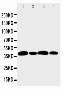
| Description | Rabbit IgG polyclonal antibody for Aurora kinase B(AURKB) detection. Tested with WB in Mouse;Rat. |
| Reconstitution | Add 0.2ml of distilled water will yield a concentration of 500ug/ml. |
| Gene ID | 20877 |
|---|---|
| Other Names | Aurora kinase B, 2.7.11.1, Aurora 1, Aurora- and IPL1-like midbody-associated protein 1, Aurora/IPL1-related kinase 2, ARK-2, Aurora-related kinase 2, STK-1, Serine/threonine-protein kinase 12, Serine/threonine-protein kinase 5, Serine/threonine-protein kinase aurora-B, Aurkb, Aik2, Aim1, Airk2, Ark2, Stk1, Stk12, Stk5 |
| Calculated MW | 39384 Da |
| Application Details | Western blot, 0.1-0.5 µg/ml, Rat, Mouse |
| Subcellular Localization | Nucleus . Chromosome . Chromosome, centromere . Cytoplasm, cytoskeleton, spindle . Midbody . Localizes on chromosome arms and inner centromeres from prophase through metaphase and then transferring to the spindle midzone and midbody from anaphase through cytokinesis. Colocalized with gamma tubulin in the mid-body (By similarity). Proper localization of the active, Thr-237-phosphorylated form during metaphase may be dependent upon interaction with SPDYC. Colocalized with SIRT2 during cytokinesis with the midbody (By similarity). . |
| Tissue Specificity | Expressed in testis, intestine and spleen. All of them are tissues that contain a large number of proliferating cells. Expressed during S phase, in a cell-cycle-dependent fashion. |
| Source | Eukaryota |
| Protein Name | Aurora kinase B |
| Contents | Each vial contains 5mg BSA, 0.9mg NaCl, 0.2mg Na2HPO4, 0.05mg Thimerosal, 0.05mg NaN3. |
| Immunogen | A synthetic peptide corresponding to a sequence at the N-terminus of mouse Aurora B(72-87aa QNKQPFTIDNFEIGRP), different from the related rat sequence by two anino acids. |
| Purification | Immunogen affinity purified. |
| Cross Reactivity | No cross reactivity with other proteins |
| Storage | At -20˚C for one year. After r˚Constitution, at 4˚C for one month. It˚Can also be aliquotted and stored frozen at -20˚C for a longer time.Avoid repeated freezing and thawing. |
| Sequence Similarities | Belongs to the protein kinase superfamily. Ser/Thr protein kinase family. Aurora subfamily. |
For Research Use Only. Not For Use In Diagnostic Procedures.
| Name | Aurkb |
|---|---|
| Function | Serine/threonine-protein kinase component of the chromosomal passenger complex (CPC), a complex that acts as a key regulator of mitosis (By similarity). The CPC complex has essential functions at the centromere in ensuring correct chromosome alignment and segregation and is required for chromatin-induced microtubule stabilization and spindle assembly (By similarity). Involved in the bipolar attachment of spindle microtubules to kinetochores and is a key regulator for the onset of cytokinesis during mitosis (By similarity). Required for central/midzone spindle assembly and cleavage furrow formation (By similarity). Key component of the cytokinesis checkpoint, a process required to delay abscission to prevent both premature resolution of intercellular chromosome bridges and accumulation of DNA damage: phosphorylates CHMP4C, leading to retain abscission-competent VPS4 (VPS4A and/or VPS4B) at the midbody ring until abscission checkpoint signaling is terminated at late cytokinesis (By similarity). AURKB phosphorylates the CPC complex subunits BIRC5/survivin, CDCA8/borealin and INCENP (By similarity). Phosphorylation of INCENP leads to increased AURKB activity (By similarity). Other known AURKB substrates involved in centromeric functions and mitosis are CENPA, DES/desmin, GPAF, KIF2C, NSUN2, RACGAP1, SEPTIN1, VIM/vimentin, HASPIN, and histone H3 (By similarity). A positive feedback loop involving HASPIN and AURKB contributes to localization of CPC to centromeres (By similarity). Phosphorylation of VIM controls vimentin filament segregation in cytokinetic process, whereas histone H3 is phosphorylated at 'Ser-10' and 'Ser-28' during mitosis (H3S10ph and H3S28ph, respectively) (PubMed:11784863). AURKB is also required for kinetochore localization of BUB1 and SGO1 (By similarity). Phosphorylation of p53/TP53 negatively regulates its transcriptional activity (By similarity). Key regulator of active promoters in resting B- and T-lymphocytes: acts by mediating phosphorylation of H3S28ph at active promoters in resting B- cells, inhibiting RNF2/RING1B-mediated ubiquitination of histone H2A and enhancing binding and activity of the USP16 deubiquitinase at transcribed genes (PubMed:24034696). Acts as an inhibitor of CGAS during mitosis: catalyzes phosphorylation of the N-terminus of CGAS during the G2-M transition, blocking CGAS liquid phase separation and activation, and thereby preventing CGAS-induced autoimmunity (By similarity). Phosphorylates KRT5 during anaphase and telophase (PubMed:29518391). Phosphorylates ATXN10 which promotes phosphorylation of ATXN10 by PLK1 and may play a role in the regulation of cytokinesis and stimulating the proteasomal degradation of ATXN10 (By similarity). |
| Cellular Location | Nucleus {ECO:0000250|UniProtKB:Q96GD4}. Chromosome {ECO:0000250|UniProtKB:Q96GD4}. Chromosome, centromere {ECO:0000250|UniProtKB:Q96GD4}. Chromosome, centromere, kinetochore {ECO:0000250|UniProtKB:Q96GD4}. Cytoplasm, cytoskeleton, spindle {ECO:0000250|UniProtKB:Q96GD4}. Midbody {ECO:0000250|UniProtKB:Q96GD4} Note=Localizes on chromosome arms and inner centromeres from prophase through metaphase and then transferring to the spindle midzone and midbody from anaphase through cytokinesis. Colocalized with gamma tubulin in the midbody. Proper localization of the active, Thr-237- phosphorylated form during metaphase may be dependent upon interaction with SPDYC. Colocalized with SIRT2 during cytokinesis with the midbody Localization (and probably targeting of the CPC) to the inner centromere occurs predominantly in regions with overlapping mitosis- specific histone phosphorylations H3pT3 and H2ApT12 {ECO:0000250|UniProtKB:Q96GD4} |
| Tissue Location | Expressed in testis, intestine and spleen. All of them are tissues that contain a large number of proliferating cells Expressed during S phase, in a cell-cycle-dependent fashion |
Provided below are standard protocols that you may find useful for product applications.
BACKGROUND
AURKB(aurora kinase B) also called Aik2, AIM-1, ARK2, AurB, STK12, IPL1, PPP1R48 or STK5, localizes to microtubules near kinetochores, specifically to the specialized microtubules called K-fibers. Cell cycle and Northern blot analyses showed that STK12 is expressed in the S phase and persistently thereafter. Western blot analysis indicated that STK12 is localized in the midbodies during anaphase. Northern blot analysis detected strong expression of a 1.5-kb STK12 transcript in thymus, with weaker expression in small intestine, testis, colon, spleen, and brain. The AURKB gene is mapped on 17p13.1. Examination of the role of both kinases in the phosphorylation of CENPA revealed that the reaction is mediated sequentially by AURKA and AURKB in early mitosis. EB1 overexpression enhanced AURKB kinase activity, and knockdown of EB1 with small interfering RNA impaired AURKB activity. EB1 protected AURKB from dephosphorylation/inactivation by protein phosphatase-2A by blocking binding of PP2A to AURKB.Â

终于等到您。ABCEPTA(百远生物)抗体产品。
点击下方“我要评价 ”按钮提交您的反馈信息,您的反馈和评价是我们最宝贵的财富之一,
我们将在1-3个工作日内处理您的反馈信息。
如有疑问,联系:0512-88856768 tech-china@abcepta.com.


