


 癌症的基本特征包括细胞增殖、血管生成、迁移、凋亡逃避机制和细胞永生等。找到癌症发生过程中这些通路的关键标记物和对应的抗体用于检测至关重要。
 为您推荐一个泛素化位点预测神器——泛素化分析工具,可以为您的蛋白的泛素化位点作出预测和评分。
 细胞自噬受体图形绘图工具为你的蛋白的细胞受体结合位点作出预测和评分,识别结合到自噬通路中的蛋白是非常重要的,便于让我们理解自噬在正常生理、病理过程中的作用,如发育、细胞分化、神经退化性疾病、压力条件下、感染和癌症。
Anti-ITGB1 Picoband Antibody
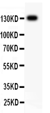
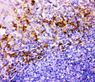
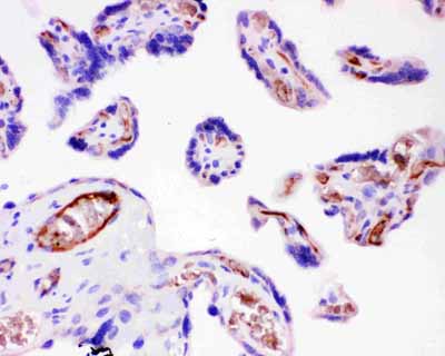
- 产品详情
- 实验流程
- 背景知识
Application 
| WB, IHC-P |
|---|---|
| Primary Accession | P05556 |
| Host | Rabbit |
| Reactivity | Human |
| Clonality | Polyclonal |
| Format | Lyophilized |
| Description | Rabbit IgG polyclonal antibody for Integrin beta-1(ITGB1) detection. Tested with WB, IHC-P in Human. |
| Reconstitution | Add 0.2ml of distilled water will yield a concentration of 500ug/ml. |
| Gene ID | 3688 |
|---|---|
| Other Names | Integrin beta-1, Fibronectin receptor subunit beta, Glycoprotein IIa, GPIIA, VLA-4 subunit beta, CD29, ITGB1, FNRB, MDF2, MSK12 |
| Calculated MW | 88415 Da |
| Application Details | Immunohistochemistry(Paraffin-embedded Section), 0.5-1 µg/ml, Human, By Heat Western blot, 0.1-0.5 µg/ml, Human |
| Subcellular Localization | Cell membrane ; Single-pass type I membrane protein . Cell projection, invadopodium membrane ; Single-pass type I membrane protein . Cell projection, ruffle membrane ; Single- pass type I membrane protein . Recycling endosome. Melanosome. Cleavage furrow. Cell projection, lamellipodium. Cell projection, ruffle. Isoform 2 does not localize to focal adhesions. Highly enriched in stage I melanosomes. Located on plasma membrane of neuroblastoma NMB7 cells. In a lung cancer cell line, in prometaphase and metaphase, localizes diffusely at the membrane and in a few intracellular vesicles. In early telophase, detected mainly on the matrix-facing side of the cells. By mid- telophase, concentrated to the ingressing cleavage furrow, mainly to the basal side of the furrow. In late telophase, concentrated to the extending protrusions formed at the opposite ends of the spreading daughter cells, in vesicles at the base of the lamellipodia formed by the separating daughter cells. Colocalizes with ITGB1BP1 and metastatic suppressor protein NME2 at the edge or peripheral ruffles and lamellipodia during the early stages of cell spreading on fibronectin or collagen. Translocates from peripheral focal adhesions sites to fibrillar adhesions in a ITGB1BP1-dependent manner. Enriched preferentially at invadopodia, cell membrane protrusions that correspond to sites of cell invasion, in a collagen-dependent manner. Localized at plasma and ruffle membranes in a collagen-independent manner. . |
| Tissue Specificity | Isoform 1 is widely expressed, other isoforms are generally coexpressed with a more restricted distribution. Isoform 2 is expressed in skin, liver, skeletal muscle, cardiac muscle, placenta, umbilical vein endothelial cells, neuroblastoma cells, lymphoma cells, hepatoma cells and astrocytoma cells. Isoform 3 and isoform 4 are expressed in muscle, kidney, liver, placenta, cervical epithelium, umbilical vein endothelial cells, fibroblast cells, embryonal kidney cells, platelets and several blood cell lines. Isoform 4, rather than isoform 3, is selectively expressed in peripheral T-cells. Isoform 3 is expressed in non- proliferating and differentiated prostate gland epithelial cells and in platelets, on the surface of erythroleukemia cells and in various hematopoietic cell lines. Isoform 5 is expressed specifically in striated muscle (skeletal and cardiac muscle). . |
| Source | Eukaryota |
| Protein Name | Integrin beta-1 |
| Contents | Each vial contains 5mg BSA, 0.9mg NaCl, 0.2mg Na2HPO4, 0.05mg NaN3. |
| Immunogen | E.coli-derived human ITGB1 recombinant protein (Position: N527-D728). Human ITGB1 shares 91% and 88% amino acid (aa) sequences identity with mouse and rat ITGB1, respectively. |
| Purification | Immunogen affinity purified. |
| Cross Reactivity | No cross reactivity with other proteins |
| Storage | At -20˚C for one year. After r˚Constitution, at 4˚C for one month. It˚Can also be aliquotted and stored frozen at -20˚C for a longer time.Avoid repeated freezing and thawing. |
| Sequence Similarities | Belongs to the integrin beta chain family. |
For Research Use Only. Not For Use In Diagnostic Procedures.
| Name | ITGB1 (HGNC:6153) |
|---|---|
| Synonyms | FNRB, MDF2, MSK12 |
| Function | Integrins alpha-1/beta-1, alpha-2/beta-1, alpha-10/beta-1 and alpha-11/beta-1 are receptors for collagen. Integrins alpha-1/beta-1 and alpha-2/beta-2 recognize the proline-hydroxylated sequence G-F-P-G- E-R in collagen. Integrins alpha-2/beta-1, alpha-3/beta-1, alpha- 4/beta-1, alpha-5/beta-1, alpha-8/beta-1, alpha-10/beta-1, alpha- 11/beta-1 and alpha-V/beta-1 are receptors for fibronectin. Alpha- 4/beta-1 recognizes one or more domains within the alternatively spliced CS-1 and CS-5 regions of fibronectin. Integrin alpha-5/beta-1 is a receptor for fibrinogen. Integrin alpha-1/beta-1, alpha-2/beta-1, alpha-6/beta-1 and alpha-7/beta-1 are receptors for lamimin. Integrin alpha-6/beta-1 (ITGA6:ITGB1) is present in oocytes and is involved in sperm-egg fusion (By similarity). Integrin alpha-4/beta-1 is a receptor for VCAM1. It recognizes the sequence Q-I-D-S in VCAM1. Integrin alpha- 9/beta-1 is a receptor for VCAM1, cytotactin and osteopontin. It recognizes the sequence A-E-I-D-G-I-E-L in cytotactin. Integrin alpha- 3/beta-1 is a receptor for epiligrin, thrombospondin and CSPG4. Alpha- 3/beta-1 may mediate with LGALS3 the stimulation by CSPG4 of endothelial cells migration. Integrin alpha-V/beta-1 is a receptor for vitronectin. Beta-1 integrins recognize the sequence R-G-D in a wide array of ligands. When associated with alpha-7 integrin, regulates cell adhesion and laminin matrix deposition. Involved in promoting endothelial cell motility and angiogenesis. Involved in osteoblast compaction through the fibronectin fibrillogenesis cell-mediated matrix assembly process and the formation of mineralized bone nodules. May be involved in up-regulation of the activity of kinases such as PKC via binding to KRT1. Together with KRT1 and RACK1, serves as a platform for SRC activation or inactivation. Plays a mechanistic adhesive role during telophase, required for the successful completion of cytokinesis. Integrin alpha-3/beta-1 provides a docking site for FAP (seprase) at invadopodia plasma membranes in a collagen-dependent manner and hence may participate in the adhesion, formation of invadopodia and matrix degradation processes, promoting cell invasion. ITGA4:ITGB1 binds to fractalkine (CX3CL1) and may act as its coreceptor in CX3CR1-dependent fractalkine signaling (PubMed:23125415, PubMed:24789099). ITGA4:ITGB1 and ITGA5:ITGB1 bind to PLA2G2A via a site (site 2) which is distinct from the classical ligand-binding site (site 1) and this induces integrin conformational changes and enhanced ligand binding to site 1 (PubMed:18635536, PubMed:25398877). ITGA5:ITGB1 acts as a receptor for fibrillin-1 (FBN1) and mediates R-G- D-dependent cell adhesion to FBN1 (PubMed:12807887, PubMed:17158881). ITGA5:ITGB1 acts as a receptor for fibronectin FN1 and mediates R-G-D- dependent cell adhesion to FN1 (PubMed:33962943). ITGA5:ITGB1 is a receptor for IL1B and binding is essential for IL1B signaling (PubMed:29030430). ITGA5:ITGB3 is a receptor for soluble CD40LG and is required for CD40/CD40LG signaling (PubMed:31331973). ITGA5:ITGB1 acts as a receptor for SNED1 and mediates R-G-D-dependent cell adhesion to SNED1 (PubMed:39713860). Plays an important role in myoblast differentiation and fusion during skeletal myogenesis (By similarity). ITGA9:ITGB1 may play a crucial role in SVEP1/polydom-mediated myoblast cell adhesion (By similarity). Integrins ITGA9:ITGB1 and ITGA4:ITGB1 repress PRKCA-mediated L-type voltage-gated channel Ca(2+) influx and ROCK-mediated calcium sensitivity in vascular smooth muscle cells via their interaction with SVEP1, thereby inhibit vasocontraction (PubMed:35802072). |
| Cellular Location | Cell membrane; Single-pass type I membrane protein. Cell projection, invadopodium membrane; Single-pass type I membrane protein. Cell projection, ruffle membrane; Single-pass type I membrane protein. Recycling endosome. Melanosome. Cleavage furrow. Cell projection, lamellipodium. Cell junction, focal adhesion. Note=Highly enriched in stage I melanosomes. Located on plasma membrane of neuroblastoma NMB7 cells. In a lung cancer cell line, in prometaphase and metaphase, localizes diffusely at the membrane and in a few intracellular vesicles. In early telophase, detected mainly on the matrix-facing side of the cells. By mid-telophase, concentrated to the ingressing cleavage furrow, mainly to the basal side of the furrow. In late telophase, concentrated to the extending protrusions formed at the opposite ends of the spreading daughter cells, in vesicles at the base of the lamellipodia formed by the separating daughter cells Colocalizes with ITGB1BP1 and metastatic suppressor protein NME2 at the edge or peripheral ruffles and lamellipodia during the early stages of cell spreading on fibronectin or collagen. Translocates from peripheral focal adhesions sites to fibrillar adhesions in a ITGB1BP1-dependent manner. Enriched preferentially at invadopodia, cell membrane protrusions that correspond to sites of cell invasion, in a collagen- dependent manner. Localized at plasma and ruffle membranes in a collagen-independent manner. [Isoform 5]: Cell membrane, sarcolemma {ECO:0000250|UniProtKB:P09055}. Cell junction {ECO:0000250|UniProtKB:P09055}. Note=In cardiac muscle, found in costameres and intercalated disks. {ECO:0000250|UniProtKB:P09055} |
| Tissue Location | Expressed in vascular smooth muscle cells (at protein level). [Isoform 2]: Expressed in skin, liver, skeletal muscle, cardiac muscle, placenta, umbilical vein endothelial cells, neuroblastoma cells, lymphoma cells, hepatoma cells and astrocytoma cells. [Isoform 4]: Together with isoform 3, is expressed in muscle, kidney, liver, placenta, cervical epithelium, umbilical vein endothelial cells, fibroblast cells, embryonal kidney cells, platelets and several blood cell lines. Rather than isoform 3, is selectively expressed in peripheral T-cells. |
Provided below are standard protocols that you may find useful for product applications.
BACKGROUND
Integrin beta-1, also known as CD29, is a protein that in humans is encoded by the ITGB1 gene. CD29 is an integrin unit associated with very late antigen receptors. It is known to conjoin with alpha-3 subunit to create alpha3beta1 complex that reacts to such molecules as netrin-1 and reelin. This gene contains a beta subunit that appears to be analogous to band-3 of integrin. It is mapped to 10p11.22. It is found that FER mediates crosstalk between CDH2 and CD29. Integrin family members are membrane receptors involved in cell adhesion and recognition in a variety of processes including embryogenesis, hemostasis, tissue repair, immune response and metastatic diffusion of tumor cells. The protein encoded by this gene is a beta subunit.

终于等到您。ABCEPTA(百远生物)抗体产品。
点击下方“我要评价 ”按钮提交您的反馈信息,您的反馈和评价是我们最宝贵的财富之一,
我们将在1-3个工作日内处理您的反馈信息。
如有疑问,联系:0512-88856768 tech-china@abcepta.com.


