


 癌症的基本特征包括细胞增殖、血管生成、迁移、凋亡逃避机制和细胞永生等。找到癌症发生过程中这些通路的关键标记物和对应的抗体用于检测至关重要。
 为您推荐一个泛素化位点预测神器——泛素化分析工具,可以为您的蛋白的泛素化位点作出预测和评分。
 细胞自噬受体图形绘图工具为你的蛋白的细胞受体结合位点作出预测和评分,识别结合到自噬通路中的蛋白是非常重要的,便于让我们理解自噬在正常生理、病理过程中的作用,如发育、细胞分化、神经退化性疾病、压力条件下、感染和癌症。
Anti-CIB1 Picoband Antibody
- 产品详情
- 实验流程
- 背景知识
Application 
| WB |
|---|---|
| Primary Accession | Q99828 |
| Host | Rabbit |
| Reactivity | Human, Mouse, Rat |
| Clonality | Polyclonal |
| Format | Lyophilized |
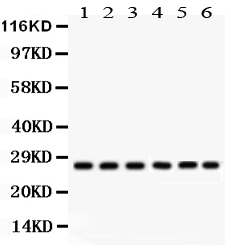
| Description | Rabbit IgG polyclonal antibody for Calcium and integrin-binding protein 1(CIB1) detection. Tested with WB in Human;Mouse;Rat. |
| Reconstitution | Add 0.2ml of distilled water will yield a concentration of 500ug/ml. |
| Gene ID | 10519 |
|---|---|
| Other Names | Calcium and integrin-binding protein 1, CIB, Calcium- and integrin-binding protein, CIBP, Calmyrin, DNA-PKcs-interacting protein, Kinase-interacting protein, KIP, SNK-interacting protein 2-28, SIP2-28, CIB1, CIB, KIP, PRKDCIP |
| Calculated MW | 21703 Da |
| Application Details | Western blot, 0.1-0.5 µg/ml, Human, Mouse, Rat |
| Subcellular Localization | Membrane; Lipid-anchor. Cell membrane, sarcolemma. Cell membrane. Apical cell membrane. Cell projection, ruffle membrane. Cell projection, filopodium tip. Cell projection, growth cone. Cell projection, lamellipodium. Cytoplasm. Cytoplasm, cytoskeleton. Cytoplasm, cytoskeleton, microtubule organizing center, centrosome. Cytoplasm, perinuclear region. Nucleus. Colocalized with PPP3R1 at the cell membrane of cardiomyocytes in the hypertrophic heart (By similarity). Colocalized with NBR1 to the perinuclear region. Colocalizes with TAS1R2 in apical regions of taste receptor cells. Colocalized with RAC3 in the perinuclear area and at the cell periphery. Colocalized with PAK1 within membrane ruffles during cell spreading upon readhesion to fibronectin. Redistributed to the cytoskeleton upon platelet aggregation. Translocates from the cytosol to the plasma membrane in a calcium-dependent manner. Colocalized with STMN2 in the cell body, neurites and growth cones of neurons. Colocalized with STMN2 to the leading edge of lamellipodia. Colocalized with PLK3 at centrosomes in ductal breast carcinoma cells. . |
| Tissue Specificity | Detected in platelets and in cell lines of megakaryocytic and erythrocytic lineages. Both isoform 1 and isoform 2 are detected in various cancer cell lines, with isoform 2 being the predominant form (at protein level). Ubiquitously expressed. . |
| Source | Eukaryota |
| Protein Name | Calcium and integrin-binding protein 1 |
| Contents | Each vial contains 5mg BSA, 0.9mg NaCl, 0.2mg Na2HPO4, 0.05mg NaN3. |
| Immunogen | E.coli-derived human CIB1 recombinant protein (Position: S6-L191). Human CIB1 shares 93.5% amino acid (aa) sequence identity with both mouse and rat CIB1. |
| Purification | Immunogen affinity purified. |
| Cross Reactivity | No cross reactivity with other proteins |
| Storage | At -20˚C for one year. After r˚Constitution, at 4˚C for one month. It˚Can also be aliquotted and stored frozen at -20˚C for a longer time.Avoid repeated freezing and thawing. |
For Research Use Only. Not For Use In Diagnostic Procedures.
| Name | CIB1 |
|---|---|
| Synonyms | CIB, KIP, PRKDCIP |
| Function | Calcium-binding protein that plays a role in the regulation of numerous cellular processes, such as cell differentiation, cell division, cell proliferation, cell migration, thrombosis, angiogenesis, cardiac hypertrophy and apoptosis. Involved in bone marrow megakaryocyte differentiation by negatively regulating thrombopoietin- mediated signaling pathway. Participates in the endomitotic cell cycle of megakaryocyte, a form of mitosis in which both karyokinesis and cytokinesis are interrupted. Plays a role in integrin signaling by negatively regulating alpha-IIb/beta3 activation in thrombin-stimulated megakaryocytes preventing platelet aggregation. Up-regulates PTK2/FAK1 activity, and is also needed for the recruitment of PTK2/FAK1 to focal adhesions; it thus appears to play an important role in focal adhesion formation. Positively regulates cell migration on fibronectin in a CDC42-dependent manner, the effect being negatively regulated by PAK1. Functions as a negative regulator of stress activated MAP kinase (MAPK) signaling pathways. Down-regulates inositol 1,4,5-trisphosphate receptor-dependent calcium signaling. Involved in sphingosine kinase SPHK1 translocation to the plasma membrane in a N-myristoylation- dependent manner preventing TNF-induced apoptosis. Regulates serine/threonine-protein kinase PLK3 activity for proper completion of cell division progression. Plays a role in microtubule (MT) dynamics during neuronal development; disrupts the MT depolymerization activity of STMN2 attenuating NGF-induced neurite outgrowth and the MT reorganization at the edge of lamellipodia. Promotes cardiomyocyte hypertrophy via activation of the calcineurin/NFAT signaling pathway. Stimulates calcineurin PPP3R1 activity by mediating its anchoring to the sarcolemma. In ischemia-induced (pathological or adaptive) angiogenesis, stimulates endothelial cell proliferation, migration and microvessel formation by activating the PAK1 and ERK1/ERK2 signaling pathway. Also promotes cancer cell survival and proliferation. May regulate cell cycle and differentiation of spermatogenic germ cells, and/or differentiation of supporting Sertoli cells. Forms a complex with TMC6/EVER1 and TMC8/EVER2 in lymphocytes and keratynocytes where CIB1 stabilizes TMC6 and TMC8 levels and reciprocally (PubMed:30068544, PubMed:32917726). |
| Cellular Location | Membrane; Lipid-anchor. Cell membrane, sarcolemma. Cell membrane. Apical cell membrane. Cell projection, ruffle membrane. Cell projection, filopodium tip. Cell projection, growth cone. Cell projection, lamellipodium. Cytoplasm. Cytoplasm, cytoskeleton. Cytoplasm, cytoskeleton, microtubule organizing center, centrosome. Cytoplasm, perinuclear region. Nucleus. Cell projection, neuron projection. Perikaryon. Note=Colocalized with PPP3R1 at the cell membrane of cardiomyocytes in the hypertrophic heart (By similarity) Colocalized with NBR1 to the perinuclear region. Colocalizes with TAS1R2 in apical regions of taste receptor cells. Colocalized with RAC3 in the perinuclear area and at the cell periphery. Colocalized with PAK1 within membrane ruffles during cell spreading upon readhesion to fibronectin. Redistributed to the cytoskeleton upon platelet aggregation. Translocates from the cytosol to the plasma membrane in a calcium-dependent manner. Colocalized with PLK3 at centrosomes in ductal breast carcinoma cells. |
| Tissue Location | Ubiquitously expressed. Expressed in the epidermis, hair follicles and keratinocytes (PubMed:30068544). Detected in platelets and in cell lines of megakaryocytic and erythrocytic lineages. Both isoform 1 and isoform 2 are detected in various cancer cell lines, with isoform 2 being the predominant form (at protein level). |
Provided below are standard protocols that you may find useful for product applications.
BACKGROUND
Calcium and integrin-binding protein 1 is a protein that in humans is encoded by the CIB1 gene. This gene encodes a member of the EF-hand domain-containing calcium-binding superfamily. And this protein interacts with many other proteins, including the platelet integrin alpha-IIb-beta-3, DNA-dependent protein kinase, presenilin-2, focal adhesion kinase, p21 activated kinase, and protein kinase D. Moreover, the encoded protein may be involved in cell survival and proliferation, and is associated with several disease states including cancer and Alzheimer's disease. Alternative splicing results in multiple transcript variants.

终于等到您。ABCEPTA(百远生物)抗体产品。
点击下方“我要评价 ”按钮提交您的反馈信息,您的反馈和评价是我们最宝贵的财富之一,
我们将在1-3个工作日内处理您的反馈信息。
如有疑问,联系:0512-88856768 tech-china@abcepta.com.


