


 癌症的基本特征包括细胞增殖、血管生成、迁移、凋亡逃避机制和细胞永生等。找到癌症发生过程中这些通路的关键标记物和对应的抗体用于检测至关重要。
 为您推荐一个泛素化位点预测神器——泛素化分析工具,可以为您的蛋白的泛素化位点作出预测和评分。
 细胞自噬受体图形绘图工具为你的蛋白的细胞受体结合位点作出预测和评分,识别结合到自噬通路中的蛋白是非常重要的,便于让我们理解自噬在正常生理、病理过程中的作用,如发育、细胞分化、神经退化性疾病、压力条件下、感染和癌症。
Anti-Cdk4 Picoband Antibody
- 产品详情
- 实验流程
- 背景知识
Application 
| WB, IHC-P |
|---|---|
| Primary Accession | P11802 |
| Host | Rabbit |
| Reactivity | Human, Mouse, Rat |
| Clonality | Polyclonal |
| Format | Lyophilized |
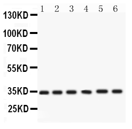
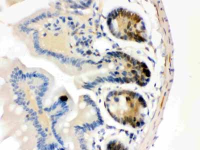
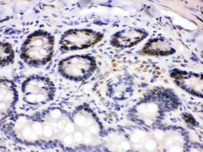
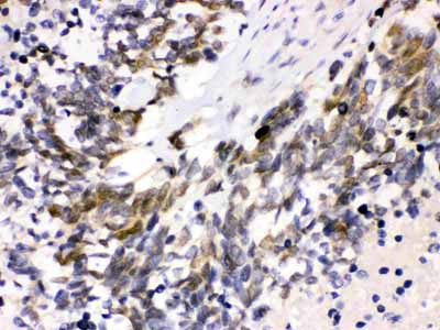
| Description | Rabbit IgG polyclonal antibody for Cyclin-dependent kinase 4(CDK4) detection. Tested with WB, IHC-P in Human;Mouse;Rat. |
| Reconstitution | Add 0.2ml of distilled water will yield a concentration of 500ug/ml. |
| Gene ID | 1019 |
|---|---|
| Other Names | Cyclin-dependent kinase 4, 2.7.11.22, Cell division protein kinase 4, PSK-J3, CDK4 |
| Calculated MW | 33730 Da |
| Application Details | Immunohistochemistry(Paraffin-embedded Section), 0.5-1 µg/ml, Human, Mouse, Rat, By Heat Western blot, 0.1-0.5 µg/ml, Human, Rat |
| Subcellular Localization | Cytoplasm. Nucleus. Membrane. Cytoplasmic when non-complexed. Forms a cyclin D-CDK4 complex in the cytoplasm as cells progress through G(1) phase. The complex accumulates on the nuclear membrane and enters the nucleus on transition from G(1) to S phase. Also present in nucleoli and heterochromatin lumps. Colocalizes with RB1 after release into the nucleus. |
| Source | Eukaryota |
| Protein Name | Cyclin-dependent kinase 4 |
| Contents | Each vial contains 5mg BSA, 0.9mg NaCl, 0.2mg Na2HPO4, 0.05mg NaN3. |
| Immunogen | E.coli-derived human Cdk4 recombinant protein (Position: G201-E303). Human Cdk4 shares 94.2% amino acid (aa) sequence identity with both mouse and rat Cdk4. |
| Purification | Immunogen affinity purified. |
| Cross Reactivity | No cross reactivity with other proteins |
| Storage | At -20˚C for one year. After r˚Constitution, at 4˚C for one month. It˚Can also be aliquotted and stored frozen at -20˚C for a longer time.Avoid repeated freezing and thawing. |
| Sequence Similarities | Belongs to the protein kinase superfamily. CMGC Ser/Thr protein kinase family. CDC2/CDKX subfamily. |
For Research Use Only. Not For Use In Diagnostic Procedures.
| Name | CDK4 {ECO:0000303|PubMed:7652577, ECO:0000312|HGNC:HGNC:1773} |
|---|---|
| Function | Ser/Thr-kinase component of cyclin D-CDK4 (DC) complexes that phosphorylate and inhibit members of the retinoblastoma (RB) protein family including RB1 and regulate the cell-cycle during G(1)/S transition. Phosphorylation of RB1 allows dissociation of the transcription factor E2F from the RB/E2F complexes and the subsequent transcription of E2F target genes which are responsible for the progression through the G(1) phase. Hypophosphorylates RB1 in early G(1) phase. Cyclin D-CDK4 complexes are major integrators of various mitogenenic and antimitogenic signals. Also phosphorylates SMAD3 in a cell-cycle-dependent manner and represses its transcriptional activity. Component of the ternary complex, cyclin D/CDK4/CDKN1B, required for nuclear translocation and activity of the cyclin D-CDK4 complex. |
| Cellular Location | Cytoplasm. Nucleus. Nucleus membrane. Note=Cytoplasmic when non-complexed Forms a cyclin D-CDK4 complex in the cytoplasm as cells progress through G(1) phase. The complex accumulates on the nuclear membrane and enters the nucleus on transition from G(1) to S phase. Also present in nucleoli and heterochromatin lumps. Colocalizes with RB1 after release into the nucleus. |
Provided below are standard protocols that you may find useful for product applications.
BACKGROUND
Cyclin-dependent kinase-4 (CDK4) is a protein-serine kinase involved in the cell cycle. Human cell division is regulated primarily at the G1-to-S or the G2-to-M boundaries within the cell cycle. The complexes formed by CDK4 and the D-type cyclins are involved in the control of cell proliferation during the G1 phase. CDK4 is inhibited by p16, also known as cyclin-dependent kinase inhibitor-2. CDK4 is mapped to 12q14. And CDK4 expression and activity are required for cytokine responsiveness in T cells.

终于等到您。ABCEPTA(百远生物)抗体产品。
点击下方“我要评价 ”按钮提交您的反馈信息,您的反馈和评价是我们最宝贵的财富之一,
我们将在1-3个工作日内处理您的反馈信息。
如有疑问,联系:0512-88856768 tech-china@abcepta.com.


