


 癌症的基本特征包括细胞增殖、血管生成、迁移、凋亡逃避机制和细胞永生等。找到癌症发生过程中这些通路的关键标记物和对应的抗体用于检测至关重要。
 为您推荐一个泛素化位点预测神器——泛素化分析工具,可以为您的蛋白的泛素化位点作出预测和评分。
 细胞自噬受体图形绘图工具为你的蛋白的细胞受体结合位点作出预测和评分,识别结合到自噬通路中的蛋白是非常重要的,便于让我们理解自噬在正常生理、病理过程中的作用,如发育、细胞分化、神经退化性疾病、压力条件下、感染和癌症。
Anti-Kv1.2 Picoband Antibody
- 产品详情
- 实验流程
- 背景知识
Application 
| WB |
|---|---|
| Primary Accession | P16389 |
| Host | Rabbit |
| Reactivity | Human, Mouse, Rat |
| Clonality | Polyclonal |
| Format | Lyophilized |
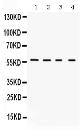
| Description | Rabbit IgG polyclonal antibody for Potassium voltage-gated channel subfamily A member 2(KCNA2) detection. Tested with WB in Human;Mouse;Rat. |
| Reconstitution | Add 0.2ml of distilled water will yield a concentration of 500ug/ml. |
| Gene ID | 3737 |
|---|---|
| Other Names | Potassium voltage-gated channel subfamily A member 2, NGK1, Voltage-gated K(+) channel HuKIV, Voltage-gated potassium channel HBK5, Voltage-gated potassium channel subunit Kv1.2, KCNA2 |
| Calculated MW | 56717 Da |
| Application Details | Western blot, 0.1-0.5 µg/ml, Mouse, Rat, Human |
| Subcellular Localization | Cell membrane ; Multi- pass membrane protein . Membrane . Cell projection, axon . Cell junction, synapse . Endoplasmic reticulum membrane . Cell projection, lamellipodium membrane . Cell junction, synapse, synaptosome . Cell junction, synapse, presynaptic cell membrane . Cell projection, dendrite . KCNA2 by itself is detected both at the endoplasmic reticulum and at the cell membrane. Coexpression with KCNA4 or with beta subunits promotes expression at the cell membrane. Coexpression with KCNA1 inhibits cell surface expression. . |
| Tissue Specificity | Detected in brain cortex (PubMed:16473933). Detected in peroneal nerve in the juxtaparanodal regions of the node of Ranvier; expression is decreased in patients with diabetes mellitus that suffer from axonal neuropathy (PubMed:22649228). Detected in paranodal and juxtanodal zones in myelinated spinal cord (at protein level) (PubMed:11086297). . |
| Source | Eukaryota |
| Protein Name | Potassium voltage-gated channel subfamily A member 2 |
| Contents | Each vial contains 5mg BSA, 0.9mg NaCl, 0.2mg Na2HPO4, 0.05mg NaN3. |
| Immunogen | A synthetic peptide corresponding to a sequence at the C-terminus of human Kv1.2 (466-499aa NNSNEDFREENLKTANCTLANTNYVNITKMLTDV), identical to the related mouse and rat sequences. |
| Purification | Immunogen affinity purified. |
| Cross Reactivity | No cross reactivity with other proteins |
| Storage | At -20˚C for one year. After r˚Constitution, at 4˚C for one month. It˚Can also be aliquotted and stored frozen at -20˚C for a longer time.Avoid repeated freezing and thawing. |
For Research Use Only. Not For Use In Diagnostic Procedures.
| Name | KCNA2 |
|---|---|
| Function | Voltage-gated potassium channel that mediates transmembrane potassium transport in excitable membranes, primarily in the brain and the central nervous system, but also in the cardiovascular system. Prevents aberrant action potential firing and regulates neuronal output. Forms tetrameric potassium-selective channels through which potassium ions pass in accordance with their electrochemical gradient. The channel alternates between opened and closed conformations in response to the voltage difference across the membrane (PubMed:11211111, PubMed:19912772, PubMed:23769686, PubMed:8495559). Can form functional homotetrameric channels and heterotetrameric channels that contain variable proportions of KCNA1, KCNA2, KCNA4, KCNA5, KCNA6, KCNA7, and possibly other family members as well; channel properties depend on the type of alpha subunits that are part of the channel (PubMed:20220134, PubMed:8495559). Channel properties are modulated by cytoplasmic beta subunits that regulate the subcellular location of the alpha subunits and promote rapid inactivation of delayed rectifier potassium channels. In vivo, membranes probably contain a mixture of heteromeric potassium channel complexes, making it difficult to assign currents observed in intact tissues to any particular potassium channel family member. Homotetrameric KCNA2 forms a delayed-rectifier potassium channel that opens in response to membrane depolarization, followed by slow spontaneous channel closure (PubMed:19912772, PubMed:23769686). In contrast, a heteromultimer formed by KCNA2 and KCNA4 shows rapid inactivation (PubMed:8495559). Regulates neuronal excitability and plays a role as pacemaker in the regulation of neuronal action potentials (By similarity). KCNA2- containing channels play a presynaptic role and prevent hyperexcitability and aberrant action potential firing (By similarity). Response to toxins that are selective for KCNA2-containing potassium channels suggests that in Purkinje cells, dendritic subthreshold KCNA2- containing potassium channels prevent random spontaneous calcium spikes, suppressing dendritic hyperexcitability without hindering the generation of somatic action potentials, and thereby play an important role in motor coordination (By similarity). Plays a role in the induction of long-term potentiation of neuron excitability in the CA3 layer of the hippocampus (By similarity). May function as down-stream effector for G protein-coupled receptors and inhibit GABAergic inputs to basolateral amygdala neurons (By similarity). May contribute to the regulation of neurotransmitter release, such as gamma-aminobutyric acid (GABA) (By similarity). Contributes to the regulation of the axonal release of the neurotransmitter dopamine (By similarity). Reduced KCNA2 expression plays a role in the perception of neuropathic pain after peripheral nerve injury, but not acute pain (By similarity). Plays a role in the regulation of the time spent in non-rapid eye movement (NREM) sleep (By similarity). |
| Cellular Location | Cell membrane; Multi-pass membrane protein {ECO:0000250|UniProtKB:P63142, ECO:0000305}. Membrane {ECO:0000250|UniProtKB:P63142}. Cell projection, axon. Synapse {ECO:0000250|UniProtKB:P63142} Endoplasmic reticulum membrane {ECO:0000250|UniProtKB:P63142}. Cell projection, lamellipodium membrane {ECO:0000250|UniProtKB:P63142} Synapse, synaptosome {ECO:0000250|UniProtKB:P63141}. Presynaptic cell membrane {ECO:0000250|UniProtKB:P63141}. Cell projection, dendrite {ECO:0000250|UniProtKB:P63141}. Cell junction, paranodal septate junction {ECO:0000250|UniProtKB:P63141}. Note=KCNA2 by itself is detected both at the endoplasmic reticulum and at the cell membrane Coexpression with KCNA4 or with beta subunits promotes expression at the cell membrane. Coexpression with KCNA1 inhibits cell surface expression. In myelinated peripheral axons, clustered in the juxtaparadonal region and at an internodal line located along the mesaxon and below the Schmidt-Lanterman incisures (By similarity) {ECO:0000250|UniProtKB:P63141, ECO:0000250|UniProtKB:P63142} |
| Tissue Location | Detected in brain cortex (PubMed:16473933). Detected in peroneal nerve in the juxtaparanodal regions of the node of Ranvier; expression is decreased in patients with diabetes mellitus that suffer from axonal neuropathy (PubMed:22649228). Detected in paranodal and juxtanodal zones in myelinated spinal cord (at protein level) (PubMed:11086297). |
Provided below are standard protocols that you may find useful for product applications.
BACKGROUND
Potassium voltage-gated channel subfamily A member 2, also known as Kv1.2, is a protein that in humans is encoded by the KCNA2 gene. Potassium channels represent the most complex class of voltage-gated ion channels from both functional and structural standpoints. Their diverse functions include regulating neurotransmitter release, heart rate, insulin secretion, neuronal excitability, epithelial electrolyte transport, smooth muscle contraction, and cell volume. Four sequence-related potassium channel genes - shaker, shaw, shab, and shal - have been identified in Drosophila, and each has been shown to have human homolog(s). This gene encodes a member of the potassium channel, voltage-gated, shaker-related subfamily. This member contains six membrane-spanning domains with a shaker-type repeat in the fourth segment. It belongs to the delayed rectifier class, members of which allow nerve cells to efficiently repolarize following an action potential. The coding region of this gene is intronless, and the gene is clustered with genes KCNA3 and KCNA10 on chromosome 1.

终于等到您。ABCEPTA(百远生物)抗体产品。
点击下方“我要评价 ”按钮提交您的反馈信息,您的反馈和评价是我们最宝贵的财富之一,
我们将在1-3个工作日内处理您的反馈信息。
如有疑问,联系:0512-88856768 tech-china@abcepta.com.


