


 癌症的基本特征包括细胞增殖、血管生成、迁移、凋亡逃避机制和细胞永生等。找到癌症发生过程中这些通路的关键标记物和对应的抗体用于检测至关重要。
 为您推荐一个泛素化位点预测神器——泛素化分析工具,可以为您的蛋白的泛素化位点作出预测和评分。
 细胞自噬受体图形绘图工具为你的蛋白的细胞受体结合位点作出预测和评分,识别结合到自噬通路中的蛋白是非常重要的,便于让我们理解自噬在正常生理、病理过程中的作用,如发育、细胞分化、神经退化性疾病、压力条件下、感染和癌症。
Anti-FE65 Antibody
- 产品详情
- 实验流程
- 背景知识
Application 
| WB |
|---|---|
| Primary Accession | O00213 |
| Host | Rabbit |
| Reactivity | Human, Mouse |
| Clonality | Polyclonal |
| Format | Lyophilized |
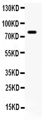
| Description | Rabbit IgG polyclonal antibody for Amyloid beta A4 precursor protein-binding family B member 1(APBB1) detection. Tested with WB in Human;Mouse. |
| Reconstitution | Add 0.2ml of distilled water will yield a concentration of 500ug/ml. |
| Gene ID | 322 |
|---|---|
| Other Names | Amyloid beta A4 precursor protein-binding family B member 1, Protein Fe65, APBB1, FE65, RIR |
| Calculated MW | 77244 Da |
| Application Details | Western blot, 0.1-0.5 µg/ml, Human, Mouse |
| Subcellular Localization | Cell membrane. Cytoplasm. Nucleus. Cell projection, growth cone . Nucleus speckle. Colocalizes with TSHZ3 in axonal growth cone (By similarity). In normal conditions, it mainly localizes to the cytoplasm, while a small fraction is tethered to the cell membrane via its interaction with APP. Following exposure to DNA damaging agents, it is released from cell membrane and translocates to the nucleus. Nuclear translocation is under the regulation of APP. Colocalizes with TSHZ3 in the nucleus. Colocalizes with NEK6 at the nuclear speckles. Phosphorylation at Ser-610 by SGK1 promotes its localization to the nucleus (By similarity). . |
| Tissue Specificity | Highly expressed in brain; strongly reduced in post-mortem elderly subjects with Alzheimer disease. . |
| Source | Eukaryota |
| Protein Name | Amyloid beta A4 precursor protein-binding family B member 1 |
| Contents | Each vial contains 5mg BSA, 0.9mg NaCl, 0.2mg Na2HPO4, 0.05mg NaN3. |
| Immunogen | E.coli-derived human FE65 recombinant protein (Position: Q295-A613). Human FE65 shares 95%and 96% amino acid (aa) sequence identity with mouse and rat FE65, respectively. |
| Purification | Immunogen affinity purified. |
| Cross Reactivity | No cross reactivity with other proteins |
| Storage | At -20˚C for one year. After r˚Constitution, at 4˚C for one month. It˚Can also be aliquotted and stored frozen at -20˚C for a longer time.Avoid repeated freezing and thawing. |
| Sequence Similarities | Contains 2 PID domains. |
For Research Use Only. Not For Use In Diagnostic Procedures.
| Name | APBB1 (HGNC:581) |
|---|---|
| Function | Transcription coregulator that can have both coactivator and corepressor functions (PubMed:15031292, PubMed:18468999, PubMed:18922798, PubMed:25342469, PubMed:33938178). Adapter protein that forms a transcriptionally active complex with the gamma-secretase- derived amyloid precursor protein (APP) intracellular domain (PubMed:15031292, PubMed:18468999, PubMed:18922798, PubMed:25342469). Plays a central role in the response to DNA damage by translocating to the nucleus and inducing apoptosis (PubMed:15031292, PubMed:18468999, PubMed:18922798, PubMed:25342469). May act by specifically recognizing and binding histone H2AX phosphorylated on 'Tyr-142' (H2AXY142ph) at double-strand breaks (DSBs), recruiting other pro-apoptosis factors such as MAPK8/JNK1 (PubMed:19234442). Required for histone H4 acetylation at double-strand breaks (DSBs) (PubMed:19234442). Its ability to specifically bind modified histones and chromatin modifying enzymes such as KAT5/TIP60, probably explains its transcription activation activity (PubMed:33938178). Functions in association with TSHZ3, SET and HDAC factors as a transcriptional repressor, that inhibits the expression of CASP4 (PubMed:19343227). Associates with chromatin in a region surrounding the CASP4 transcriptional start site(s) (PubMed:19343227). Involved in hippocampal neurite branching and neuromuscular junction formation, as a result plays a role in spatial memory functioning (By similarity). Plays a role in the maintenance of lens transparency (By similarity). May play a role in muscle cell strength (By similarity). Acts as a molecular adapter that functions in neurite outgrowth by activating the RAC1-ARF6 axis upon insulin treatment (PubMed:36250347). |
| Cellular Location | Cell membrane. Cytoplasm. Nucleus. Cell projection, growth cone {ECO:0000250|UniProtKB:P46933}. Nucleus speckle. Note=Colocalizes with TSHZ3 in axonal growth cone (By similarity). Colocalizes with TSHZ3 in the nucleus (PubMed:19343227). In normal conditions, it mainly localizes to the cytoplasm, while a small fraction is tethered to the cell membrane via its interaction with APP (PubMed:18468999). Following exposure to DNA damaging agents, it is released from cell membrane and translocates to the nucleus (PubMed:18468999). Nuclear translocation is under the regulation of APP (PubMed:18468999). Colocalizes with NEK6 at the nuclear speckles (PubMed:17512906). Phosphorylation at Ser-610 by SGK1 promotes its localization to the nucleus (By similarity) {ECO:0000250|UniProtKB:P46933, ECO:0000269|PubMed:17512906, ECO:0000269|PubMed:18468999, ECO:0000269|PubMed:19343227} |
| Tissue Location | Highly expressed in brain; strongly reduced in post-mortem elderly subjects with Alzheimer disease |
Provided below are standard protocols that you may find useful for product applications.
BACKGROUND
APBB1 is also known as RIR or FE65. The protein encoded by this gene is a member of the Fe65 protein family. It is an adaptor protein localized in the nucleus. It interacts with the Alzheimer's disease amyloid precursor protein (APP), transcription factor CP2/LSF/LBP1 and the low-density lipoprotein receptor-related protein. APP functions as a cytosolic anchoring site that can prevent the gene product's nuclear translocation. This encoded protein could play an important role in the pathogenesis of Alzheimer's disease. It is thought to regulate transcription. Also it is observed to block cell cycle progression by downregulating thymidylate synthase expression. Multiple alternatively spliced transcript variants encoding different isoforms have been described for this gene.

终于等到您。ABCEPTA(百远生物)抗体产品。
点击下方“我要评价 ”按钮提交您的反馈信息,您的反馈和评价是我们最宝贵的财富之一,
我们将在1-3个工作日内处理您的反馈信息。
如有疑问,联系:0512-88856768 tech-china@abcepta.com.


