注册 | 登录
点击这里给我发消息
所有产品
  • 所有产品
  • 一抗
  • 裂解液
>   首页   >   产品   >   一抗   >   信号转导   >   Anti-Vitamin D Binding protein Picoband Antibody   
Anti-Vitamin D Binding protein Picoband Antibody
     
  • 1 - Anti-Vitamin D Binding protein Picoband Antibody ABO13011
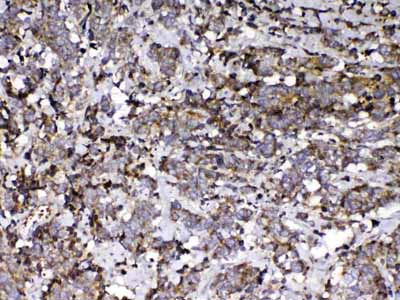
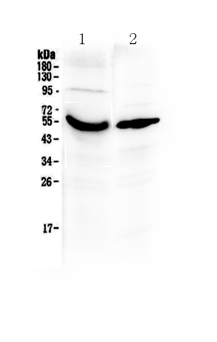
    Figure 1. Western blot analysis of Vitamin D Binding protein using anti-Vitamin D Binding protein antibody (ABO13011). Electrophoresis was performed on a 5-20% SDS-PAGE gel at 70V (Stacking gel) / 90V (Resolving gel) for 2-3 hours. The sample well of each lane was loaded with 50ug of sample under reducing conditions. Lane 1: human placenta tissue lysates,Lane 2: human A431 whole cell lysates. After Electrophoresis, proteins were transferred to a Nitrocellulose membrane at 150mA for 50-90 minutes. Blocked the membrane with 5% Non-fat Milk/ TBS for 1.5 hour at RT. The membrane was incubated with rabbit anti-Vitamin D Binding protein antigen affinity purified polyclonal antibody (Catalog # ABO13011) at 0.5 ug/mL overnight at 4℃, then washed with TBS-0.1%Tween 3 times with 5 minutes each and probed with a goat anti-rabbit IgG-HRP secondary antibody at a dilution of 1:10000 for 1.5 hour at RT. The signal is developed using an Enhanced Chemiluminescent detection (ECL) kit with Tanon 5200 system. A specific band was detected for Vitamin D Binding protein at approximately 53KD. The expected band size for Vitamin D Binding protein is at 53KD.
  • 2 - Anti-Vitamin D Binding protein Picoband Antibody ABO13011
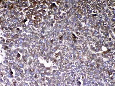
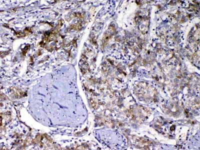
    Figure 2. IHC analysis of Vitamin D Binding protein using anti-Vitamin D Binding protein antibody (ABO13011).Vitamin D Binding protein was detected in paraffin-embedded section of human liver cancer tissue. Heat mediated antigen retrieval was performed in citrate buffer (pH6, epitope retrieval solution) for 20 mins. The tissue section was blocked with 10% goat serum. The tissue section was then incubated with 1ug/ml rabbit anti-Vitamin D Binding protein Antibody (ABO13011) overnight at 4℃. Biotinylated goat anti-rabbit IgG was used as secondary antibody and incubated for 30 minutes at 37℃. The tissue section was developed using Strepavidin-Biotin-Complex (SABC) with DAB as the chromogen.
  • 2 - Anti-Vitamin D Binding protein Picoband Antibody ABO13011
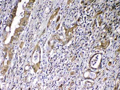
    Figure 3. IHC analysis of Vitamin D Binding protein using anti-Vitamin D Binding protein antibody (ABO13011).Vitamin D Binding protein was detected in paraffin-embedded section of human lung cancer tissue. Heat mediated antigen retrieval was performed in citrate buffer (pH6, epitope retrieval solution) for 20 mins. The tissue section was blocked with 10% goat serum. The tissue section was then incubated with 1ug/ml rabbit anti-Vitamin D Binding protein Antibody (ABO13011) overnight at 4℃. Biotinylated goat anti-rabbit IgG was used as secondary antibody and incubated for 30 minutes at 37℃. The tissue section was developed using Strepavidin-Biotin-Complex (SABC) with DAB as the chromogen.
  • 2 - Anti-Vitamin D Binding protein Picoband Antibody ABO13011
    Figure 4. IHC analysis of Vitamin D Binding protein using anti-Vitamin D Binding protein antibody (ABO13011).Vitamin D Binding protein was detected in paraffin-embedded section of mouse liver tissue. Heat mediated antigen retrieval was performed in citrate buffer (pH6, epitope retrieval solution) for 20 mins. The tissue section was blocked with 10% goat serum. The tissue section was then incubated with 1ug/ml rabbit anti-Vitamin D Binding protein Antibody (ABO13011) overnight at 4℃. Biotinylated goat anti-rabbit IgG was used as secondary antibody and incubated for 30 minutes at 37℃. The tissue section was developed using Strepavidin-Biotin-Complex (SABC) with DAB as the chromogen.
  • 2 - Anti-Vitamin D Binding protein Picoband Antibody ABO13011
    Figure 5. IHC analysis of Vitamin D Binding protein using anti-Vitamin D Binding protein antibody (ABO13011).Vitamin D Binding protein was detected in paraffin-embedded section of human rectal cancer tissue. Heat mediated antigen retrieval was performed in citrate buffer (pH6, epitope retrieval solution) for 20 mins. The tissue section was blocked with 10% goat serum. The tissue section was then incubated with 1ug/ml rabbit anti-Vitamin D Binding protein Antibody (ABO13011) overnight at 4℃. Biotinylated goat anti-rabbit IgG was used as secondary antibody and incubated for 30 minutes at 37℃. The tissue section was developed using Strepavidin-Biotin-Complex (SABC) with DAB as the chromogen.
  • 产品详情
  • 实验流程
  • 背景知识
Product Information
Application
  • Applications Legend:
  • E=ELISA
  • WB=Western Blotting
  • IHC=Immunohistochemistry
  • IHC-P=Immunohistochemistry (Paraffin)
  • IP=Immunoprecipitation
  • IF=Immunofluorescence
  • IC=Immunochemistry
  • ICC=Immunocytochemistry
  • FC=Flow Cytometry
  • DB=Dot Blot
WB, IHC-P, E
Primary Accession GC: P02774
Host Rabbit
Reactivity Human, Mouse, Rat
Clonality Polyclonal
Format Lyophilized
Description Rabbit IgG polyclonal antibody for Vitamin D Binding protein detection. Tested with WB, IHC-P, Direct ELISA in Human;Mouse;Rat.
Reconstitution Add 0.2ml of distilled water will yield a concentration of 500ug/ml.
Additional Information
Application Details Western blot, 0.1-0.5 µg/ml
Immunohistochemistry(Paraffin-embedded Section), 0.5-1 µg/ml
Direct ELISA, 0.1-0.5 µg/ml
Subcellular Localization Secreted.
Tissue Specificity Expressed in the liver. Found in plasma, ascites, cerebrospinal fluid and urine.
Contents Each vial contains 5mg BSA, 0.9mg NaCl, 0.2mg Na2HPO4, 0.05mg NaN3.
Immunogen E. coli-derived human Vitamin D Binding protein recombinant protein (Position: L17-E256).
Purification Immunogen affinity purified.
Cross Reactivity No cross reactivity with other proteins.
Storage At -20˚C for one year. After r˚Constitution, at 4˚C for one month. It˚Can also be aliquotted and stored frozen at -20˚C for a longer time. Avoid repeated freezing and thawing.

For Research Use Only. Not For Use In Diagnostic Procedures.

Protein Information
Research Areas

BACKGROUND

Vitamin D-binding protein, also/originally known as gc-globulin (group-specific component), is a protein that in humans is encoded by the GC gene.The protein encoded by this gene belongs to the albumin gene family. It is a multifunctional protein found in plasma, ascitic fluid, cerebrospinal fluid and on the surface of many cell types. It binds to vitamin D and its plasma metabolites and transports them to target tissues.

FeedBack

终于等到您。ABCEPTA(百远生物)抗体产品。
点击下方“我要评价 ”按钮提交您的反馈信息,您的反馈和评价是我们最宝贵的财富之一,
我们将在1-3个工作日内处理您的反馈信息。

如有疑问,联系:0512-88856768 tech-china@abcepta.com.


我要评价