


 癌症的基本特征包括细胞增殖、血管生成、迁移、凋亡逃避机制和细胞永生等。找到癌症发生过程中这些通路的关键标记物和对应的抗体用于检测至关重要。
 为您推荐一个泛素化位点预测神器——泛素化分析工具,可以为您的蛋白的泛素化位点作出预测和评分。
 细胞自噬受体图形绘图工具为你的蛋白的细胞受体结合位点作出预测和评分,识别结合到自噬通路中的蛋白是非常重要的,便于让我们理解自噬在正常生理、病理过程中的作用,如发育、细胞分化、神经退化性疾病、压力条件下、感染和癌症。
Anti-TMS1 PYCARD Rabbit Monoclonal Antibody
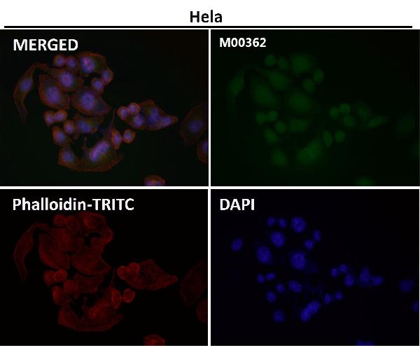
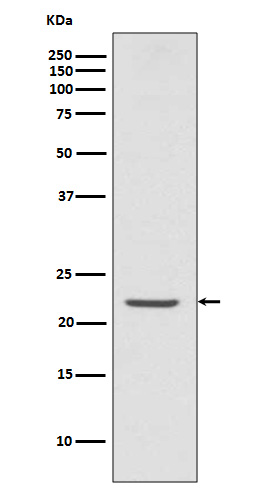
- 产品详情
- 实验流程
Application 
| WB, IF, ICC, FC |
|---|---|
| Primary Accession | Q9ULZ3 |
| Host | Rabbit |
| Isotype | Rabbit IgG |
| Reactivity | Human |
| Clonality | Monoclonal |
| Format | Liquid |
| Description | Anti-TMS1 PYCARD Rabbit Monoclonal Antibody . Tested in WB, ICC/IF, Flow Cytometry applications. This antibody reacts with Human. |
| Gene ID | 29108 |
|---|---|
| Other Names | Apoptosis-associated speck-like protein containing a CARD, hASC, Caspase recruitment domain-containing protein 5 {ECO:0000303|Ref.5}, PYD and CARD domain-containing protein {ECO:0000303|Ref.4}, Target of methylation-induced silencing 1, PYCARD {ECO:0000303|Ref.4, ECO:0000312|HGNC:HGNC:16608} |
| Calculated MW | 21627 Da |
| Application Details | WB 1:500-1:2000 ICC/IF 1:50-1:200 FC 150 |
| Subcellular Localization | Cytoplasm. Endoplasmic reticulum. Mitochondrion. Nucleus. Upstream of caspase activation, a redistribution from the cytoplasm to the aggregates occurs. These appear as hollow, perinuclear spherical, ball-like structures. Upon NLRP3 inflammasome activation redistributes to the perinuclear space localizing to endoplasmic reticulum and mitochondria. Localized primarily to the nucleus in resting monocytes/macrophages and rapidly redistributed to the cytoplasm upon pathogen infection. Localized to large cytoplasmic aggregate appearing as a speck containing AIM2, PYCARD, CASP8 and bacterial DNA after infection with Francisella tularensis (By similarity).. |
| Tissue Specificity | Widely expressed at low levels. Detected in peripheral blood leukocytes, lung, small intestine, spleen, thymus, colon and at lower levels in placenta, liver and kidney. Very low expression in skeletal muscle, heart and brain. Detected in the leukemia cell lines HL-60 and U-937, but not in Jurkat T- cell lymphoma and Daudi Burkitt's lymphoma. Detected in the melanoma cell line WM35, but not in WM793. Not detected in HeLa cervical carcinoma cells and MOLT-4 lymphocytic leukemia cells. |
| Source | Eukaryota |
| Contents | Rabbit IgG in phosphate buffered saline, pH 7.4, 150mM NaCl, 0.02% sodium azide and 50% glycerol, 0.4-0.5mg/ml BSA. |
| Clone Names | Clone: FFB-16 |
| Immunogen | A synthesized peptide derived from human TMS1 |
| Purification | Affinity-chromatography |
| Storage | Store at -20°C for one year. For short term storage and frequent use, store at 4°C for up to one month. Avoid repeated freeze-thaw cycles. |
For Research Use Only. Not For Use In Diagnostic Procedures.
| Name | PYCARD {ECO:0000303|Ref.4, ECO:0000312|HGNC:HGNC:16608} |
|---|---|
| Function | Functions as a key mediator in apoptosis and inflammation (PubMed:11103777, PubMed:12646168, PubMed:15030775, PubMed:17349957, PubMed:17599095, PubMed:19158675, PubMed:19158676, PubMed:19234215, PubMed:19494289, PubMed:21487011, PubMed:24630722, PubMed:25847972, PubMed:30674671, PubMed:34678144, PubMed:36050480). Promotes caspase- mediated apoptosis involving predominantly caspase-8 and also caspase-9 in a probable cell type-specific manner (PubMed:11103777, PubMed:12646168). Involved in activation of the mitochondrial apoptotic pathway, promotes caspase-8-dependent proteolytic maturation of BID independently of FADD in certain cell types and also mediates mitochondrial translocation of BAX and activates BAX-dependent apoptosis coupled to activation of caspase-9, -2 and -3 (PubMed:14730312, PubMed:16964285). Involved in innate immune response by acting as an integral adapter in the assembly of various inflammasomes (NLRP1, NLRP2, NLRP3, NLRP6, AIM2 and probably IFI16) which recruit and activate caspase-1 leading to processing and secretion of pro-inflammatory cytokines (PubMed:15030775, PubMed:16982856, PubMed:17349957, PubMed:17599095, PubMed:19158675, PubMed:19158676, PubMed:19234215, PubMed:21487011, PubMed:23530044, PubMed:24630722, PubMed:25847972, PubMed:29440442, PubMed:30674671, PubMed:33980849, PubMed:34678144, PubMed:34706239). Caspase-1-dependent inflammation leads to macrophage pyroptosis, a form of cell death (PubMed:24630722). The function as activating adapter in different types of inflammasomes is mediated by the pyrin and CARD domains and their homotypic interactions (PubMed:14499617, PubMed:19234215, PubMed:24630722). Clustered PYCARD nucleates the formation of caspase-1 filaments through the interaction of their respective CARD domains, acting as a platform for of caspase-1 polymerization (PubMed:24630722). In the NLRP1 and NLRC4 inflammasomes seems not be required but facilitates the processing of procaspase-1 (PubMed:17349957). In cooperation with NOD2 involved in an inflammasome activated by bacterial muramyl dipeptide leading to caspase-1 activation (PubMed:16964285). May be involved in RIGI-triggered pro-inflammatory responses and inflammasome activation (PubMed:19915568). In collaboration with AIM2 which detects cytosolic double-stranded DNA may also be involved in a caspase-1-independent cell death that involves caspase-8 (PubMed:19158675, PubMed:19158676). In adaptive immunity may be involved in maturation of dendritic cells to stimulate T-cell immunity and in cytoskeletal rearrangements coupled to chemotaxis and antigen uptake may be involved in post-transcriptional regulation of the guanine nucleotide exchange factor DOCK2; the latter function is proposed to involve the nuclear form (PubMed:22732093). Also involved in transcriptional activation of cytokines and chemokines independent of the inflammasome; this function may involve AP-1, NF-kappa-B, MAPK and caspase-8 signaling pathways (PubMed:12486103, PubMed:16585594). For regulation of NF-kappa-B activating and inhibiting functions have been reported (PubMed:12486103). Modulates NF-kappa-B induction at the level of the IKK complex by inhibiting kinase activity of CHUK and IKBK (PubMed:12486103, PubMed:16585594). Proposed to compete with RIPK2 for association with CASP1 thereby down-regulating CASP1-mediated RIPK2- dependent NF-kappa-B activation and activating interleukin-1 beta processing (PubMed:16585594). Modulates host resistance to DNA virus infection, probably by inducing the cleavage of and inactivating CGAS in presence of cytoplasmic double-stranded DNA (PubMed:28314590). |
| Cellular Location | Cytoplasm. Inflammasome. Endoplasmic reticulum. Mitochondrion. Nucleus Note=Upstream of caspase activation, a redistribution from the cytoplasm to the aggregates occurs. These appear as hollow, perinuclear spherical, ball-like structures (PubMed:11103777, PubMed:12191486, PubMed:15030775). Upon NLRP3 inflammasome activation redistributes to the perinuclear space localizing to endoplasmic reticulum and mitochondria (PubMed:12191486, PubMed:15030775). Localized primarily to the nucleus in resting monocytes/macrophages and rapidly redistributed to the cytoplasm upon pathogen infection (PubMed:19234215). Localized to large cytoplasmic aggregate appearing as a speck containing AIM2, PYCARD, CASP8 and bacterial DNA after infection with Francisella tularensis (By similarity). {ECO:0000250|UniProtKB:Q9EPB4, ECO:0000269|PubMed:11103777, ECO:0000269|PubMed:12191486, ECO:0000269|PubMed:15030775, ECO:0000269|PubMed:19234215} |
| Tissue Location | Widely expressed at low levels. Detected in peripheral blood leukocytes, lung, small intestine, spleen, thymus, colon and at lower levels in placenta, liver and kidney. Very low expression in skeletal muscle, heart and brain. Expressed in lung epithelial cells (at protein level) (PubMed:23229815). Detected in the leukemia cell lines HL-60 and U-937, but not in Jurkat T-cell lymphoma and Daudi Burkitt's lymphoma. Detected in the melanoma cell line WM35, but not in WM793. Not detected in HeLa cervical carcinoma cells and MOLT-4 lymphocytic leukemia cells. |
Research Areas
Application Protocols
Provided below are standard protocols that you may find useful for product applications.

终于等到您。ABCEPTA(百远生物)抗体产品。
点击下方“我要评价 ”按钮提交您的反馈信息,您的反馈和评价是我们最宝贵的财富之一,
我们将在1-3个工作日内处理您的反馈信息。
如有疑问,联系:0512-88856768 tech-china@abcepta.com.


