


 癌症的基本特征包括细胞增殖、血管生成、迁移、凋亡逃避机制和细胞永生等。找到癌症发生过程中这些通路的关键标记物和对应的抗体用于检测至关重要。
 为您推荐一个泛素化位点预测神器——泛素化分析工具,可以为您的蛋白的泛素化位点作出预测和评分。
 细胞自噬受体图形绘图工具为你的蛋白的细胞受体结合位点作出预测和评分,识别结合到自噬通路中的蛋白是非常重要的,便于让我们理解自噬在正常生理、病理过程中的作用,如发育、细胞分化、神经退化性疾病、压力条件下、感染和癌症。
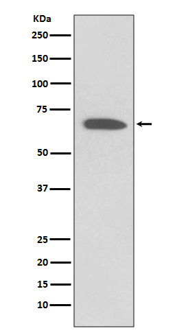
Anti-KCNA1/Kv1.1 Rabbit Monoclonal Antibody
- 产品详情
- 实验流程
Application 
| WB, IP |
|---|---|
| Primary Accession | Q09470 |
| Host | Rabbit |
| Isotype | Rabbit IgG |
| Reactivity | Human |
| Clonality | Monoclonal |
| Format | Liquid |
| Description | Anti-KCNA1/Kv1.1 Rabbit Monoclonal Antibody . Tested in WB, IP applications. This antibody reacts with Human. |
| Gene ID | 3736 |
|---|---|
| Other Names | Potassium voltage-gated channel subfamily A member 1, Voltage-gated K(+) channel HuKI, Voltage-gated potassium channel HBK1, Voltage-gated potassium channel subunit Kv1.1, KCNA1 (HGNC:6218) |
| Calculated MW | 56466 Da |
| Application Details | WB 1:5000-1:10000 IP 1:10 |
| Subcellular Localization | Cell membrane ; Multi- pass membrane protein. Membrane. Cell projection, axon. Cytoplasmic vesicle. Perikaryon. Endoplasmic reticulum. Cell projection, dendrite. Cell junction. Cell junction, synapse. Cell junction, synapse, presynaptic cell membrane. Homotetrameric KCNA1 is primarily located in the endoplasmic reticulum. Interaction with KCNA2 and KCNAB2 or with KCNA4 and KCNAB2 promotes expression at the cell membrane (By similarity). Detected at axon terminals (By similarity).. |
| Tissue Specificity | Detected adjacent to nodes of Ranvier in juxtaparanodal zones in spinal cord nerve fibers, but also in paranodal regions in some myelinated spinal cord axons (at protein level) (PubMed:11086297). Detected in the islet of Langerhans (PubMed:21483673).. |
| Source | Eukaryota |
| Contents | Rabbit IgG in phosphate buffered saline, pH 7.4, 150mM NaCl, 0.02% sodium azide and 50% glycerol, 0.4-0.5mg/ml BSA. |
| Clone Names | Clone: FFI-11 |
| Immunogen | A synthesized peptide derived from human KCNA1 |
| Purification | Affinity-chromatography |
| Storage | Store at -20°C for one year. For short term storage and frequent use, store at 4°C for up to one month. Avoid repeated freeze-thaw cycles. |
For Research Use Only. Not For Use In Diagnostic Procedures.
| Name | KCNA1 (HGNC:6218) |
|---|---|
| Function | Voltage-gated potassium channel that mediates transmembrane potassium transport in excitable membranes, primarily in the brain and the central nervous system, but also in the kidney (PubMed:19903818, PubMed:8845167). Contributes to the regulation of the membrane potential and nerve signaling, and prevents neuronal hyperexcitability (PubMed:17156368). Forms tetrameric potassium-selective channels through which potassium ions pass in accordance with their electrochemical gradient. The channel alternates between opened and closed conformations in response to the voltage difference across the membrane (PubMed:19912772). Can form functional homotetrameric channels and heterotetrameric channels that contain variable proportions of KCNA1, KCNA2, KCNA4, KCNA5, KCNA6, KCNA7, and possibly other family members as well; channel properties depend on the type of alpha subunits that are part of the channel (PubMed:12077175, PubMed:17156368). Channel properties are modulated by cytoplasmic beta subunits that regulate the subcellular location of the alpha subunits and promote rapid inactivation of delayed rectifier potassium channels (PubMed:12077175, PubMed:17156368). In vivo, membranes probably contain a mixture of heteromeric potassium channel complexes, making it difficult to assign currents observed in intact tissues to any particular potassium channel family member. Homotetrameric KCNA1 forms a delayed-rectifier potassium channel that opens in response to membrane depolarization, followed by slow spontaneous channel closure (PubMed:19307729, PubMed:19903818, PubMed:19912772, PubMed:19968958). In contrast, a heterotetrameric channel formed by KCNA1 and KCNA4 shows rapid inactivation (PubMed:17156368). Regulates neuronal excitability in hippocampus, especially in mossy fibers and medial perforant path axons, preventing neuronal hyperexcitability. Response to toxins that are selective for KCNA1, respectively for KCNA2, suggests that heteromeric potassium channels composed of both KCNA1 and KCNA2 play a role in pacemaking and regulate the output of deep cerebellar nuclear neurons (By similarity). May function as down-stream effector for G protein-coupled receptors and inhibit GABAergic inputs to basolateral amygdala neurons (By similarity). May contribute to the regulation of neurotransmitter release, such as gamma-aminobutyric acid (GABA) release (By similarity). Plays a role in regulating the generation of action potentials and preventing hyperexcitability in myelinated axons of the vagus nerve, and thereby contributes to the regulation of heart contraction (By similarity). Required for normal neuromuscular responses (PubMed:11026449, PubMed:17136396). Regulates the frequency of neuronal action potential firing in response to mechanical stimuli, and plays a role in the perception of pain caused by mechanical stimuli, but does not play a role in the perception of pain due to heat stimuli (By similarity). Required for normal responses to auditory stimuli and precise location of sound sources, but not for sound perception (By similarity). The use of toxins that block specific channels suggest that it contributes to the regulation of the axonal release of the neurotransmitter dopamine (By similarity). Required for normal postnatal brain development and normal proliferation of neuronal precursor cells in the brain (By similarity). Plays a role in the reabsorption of Mg(2+) in the distal convoluted tubules in the kidney and in magnesium ion homeostasis, probably via its effect on the membrane potential (PubMed:19307729, PubMed:23903368). |
| Cellular Location | Cell membrane; Multi-pass membrane protein. Membrane Cell projection, axon. Cytoplasmic vesicle. Perikaryon {ECO:0000250|UniProtKB:P10499}. Endoplasmic reticulum {ECO:0000250|UniProtKB:P10499}. Cell projection, dendrite {ECO:0000250|UniProtKB:P16388}. Cell junction {ECO:0000250|UniProtKB:P16388}. Synapse {ECO:0000250|UniProtKB:P16388} Presynaptic cell membrane {ECO:0000250|UniProtKB:P10499}. Presynapse {ECO:0000250|UniProtKB:P16388}. Note=Homotetrameric KCNA1 is primarily located in the endoplasmic reticulum. Interaction with KCNA2 and KCNAB2 or with KCNA4 and KCNAB2 promotes expression at the cell membrane (By similarity). {ECO:0000250|UniProtKB:P10499, ECO:0000250|UniProtKB:P16388} |
| Tissue Location | Detected adjacent to nodes of Ranvier in juxtaparanodal zones in spinal cord nerve fibers, but also in paranodal regions in some myelinated spinal cord axons (at protein level) (PubMed:11086297). Detected in the islet of Langerhans (PubMed:21483673). |
Research Areas
Application Protocols
Provided below are standard protocols that you may find useful for product applications.

终于等到您。ABCEPTA(百远生物)抗体产品。
点击下方“我要评价 ”按钮提交您的反馈信息,您的反馈和评价是我们最宝贵的财富之一,
我们将在1-3个工作日内处理您的反馈信息。
如有疑问,联系:0512-88856768 tech-china@abcepta.com.


