


 癌症的基本特征包括细胞增殖、血管生成、迁移、凋亡逃避机制和细胞永生等。找到癌症发生过程中这些通路的关键标记物和对应的抗体用于检测至关重要。
 为您推荐一个泛素化位点预测神器——泛素化分析工具,可以为您的蛋白的泛素化位点作出预测和评分。
 细胞自噬受体图形绘图工具为你的蛋白的细胞受体结合位点作出预测和评分,识别结合到自噬通路中的蛋白是非常重要的,便于让我们理解自噬在正常生理、病理过程中的作用,如发育、细胞分化、神经退化性疾病、压力条件下、感染和癌症。
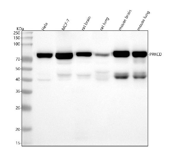
Anti-PKC delta PRKCD Rabbit Monoclonal Antibody
- 产品详情
- 实验流程
Application 
| WB, IHC, IF, ICC |
|---|---|
| Primary Accession | Q05655 |
| Host | Rabbit |
| Isotype | Rabbit IgG |
| Reactivity | Rat, Human, Mouse |
| Clonality | Monoclonal |
| Format | Liquid |
| Description | Anti-PKC delta PRKCD Rabbit Monoclonal Antibody . Tested in WB, IHC, ICC/IF applications. This antibody reacts with Human, Mouse, Rat. |
| Gene ID | 5580 |
|---|---|
| Other Names | Protein kinase C delta type, 2.7.11.13, Tyrosine-protein kinase PRKCD, 2.7.10.2, nPKC-delta, Protein kinase C delta type regulatory subunit, Protein kinase C delta type catalytic subunit, Sphingosine-dependent protein kinase-1, SDK1, PRKCD (HGNC:9399) |
| Calculated MW | 77505 Da |
| Application Details | WB 1:500-1:2000 IHC 1:50-1:200 ICC/IF 1:50-1:200 |
| Subcellular Localization | Cytoplasm. Cytoplasm, perinuclear region. Nucleus. Endoplasmic reticulum. Mitochondrion. Cell membrane; Peripheral membrane protein. |
| Source | Eukaryota |
| Contents | Rabbit IgG in phosphate buffered saline, pH 7.4, 150mM NaCl, 0.02% sodium azide and 50% glycerol, 0.4-0.5mg/ml BSA. |
| Clone Names | Clone: IHD-16 |
| Immunogen | A synthesized peptide derived from human PKC delta |
| Purification | Affinity-chromatography |
| Storage | Store at -20°C for one year. For short term storage and frequent use, store at 4°C for up to one month. Avoid repeated freeze-thaw cycles. |
For Research Use Only. Not For Use In Diagnostic Procedures.
| Name | PRKCD (HGNC:9399) |
|---|---|
| Function | Calcium-independent, phospholipid- and diacylglycerol (DAG)- dependent serine/threonine-protein kinase that plays contrasting roles in cell death and cell survival by functioning as a pro-apoptotic protein during DNA damage-induced apoptosis, but acting as an anti- apoptotic protein during cytokine receptor-initiated cell death, is involved in tumor suppression as well as survival of several cancers, is required for oxygen radical production by NADPH oxidase and acts as positive or negative regulator in platelet functional responses (PubMed:21406692, PubMed:21810427). Negatively regulates B cell proliferation and also has an important function in self-antigen induced B cell tolerance induction (By similarity). Upon DNA damage, activates the promoter of the death-promoting transcription factor BCLAF1/Btf to trigger BCLAF1-mediated p53/TP53 gene transcription and apoptosis (PubMed:21406692, PubMed:21810427). In response to oxidative stress, interact with and activate CHUK/IKKA in the nucleus, causing the phosphorylation of p53/TP53 (PubMed:21406692, PubMed:21810427). In the case of ER stress or DNA damage-induced apoptosis, can form a complex with the tyrosine-protein kinase ABL1 which trigger apoptosis independently of p53/TP53 (PubMed:21406692, PubMed:21810427). In cytosol can trigger apoptosis by activating MAPK11 or MAPK14, inhibiting AKT1 and decreasing the level of X-linked inhibitor of apoptosis protein (XIAP), whereas in nucleus induces apoptosis via the activation of MAPK8 or MAPK9. Upon ionizing radiation treatment, is required for the activation of the apoptosis regulators BAX and BAK, which trigger the mitochondrial cell death pathway. Can phosphorylate MCL1 and target it for degradation which is sufficient to trigger for BAX activation and apoptosis. Is required for the control of cell cycle progression both at G1/S and G2/M phases. Mediates phorbol 12-myristate 13-acetate (PMA)-induced inhibition of cell cycle progression at G1/S phase by up-regulating the CDK inhibitor CDKN1A/p21 and inhibiting the cyclin CCNA2 promoter activity. In response to UV irradiation can phosphorylate CDK1, which is important for the G2/M DNA damage checkpoint activation (By similarity). Can protect glioma cells from the apoptosis induced by TNFSF10/TRAIL, probably by inducing increased phosphorylation and subsequent activation of AKT1 (PubMed:15774464). Is highly expressed in a number of cancer cells and promotes cell survival and resistance against chemotherapeutic drugs by inducing cyclin D1 (CCND1) and hyperphosphorylation of RB1, and via several pro-survival pathways, including NF-kappa-B, AKT1 and MAPK1/3 (ERK1/2). Involved in antifungal immunity by mediating phosphorylation and activation of CARD9 downstream of C-type lectin receptors activation, promoting interaction between CARD9 and BCL10, followed by activation of NF- kappa-B and MAP kinase p38 pathways (By similarity). Can also act as tumor suppressor upon mitogenic stimulation with PMA or TPA. In N- formyl-methionyl-leucyl-phenylalanine (fMLP)-treated cells, is required for NCF1 (p47-phox) phosphorylation and activation of NADPH oxidase activity, and regulates TNF-elicited superoxide anion production in neutrophils, by direct phosphorylation and activation of NCF1 or indirectly through MAPK1/3 (ERK1/2) signaling pathways (PubMed:19801500). May also play a role in the regulation of NADPH oxidase activity in eosinophil after stimulation with IL5, leukotriene B4 or PMA (PubMed:11748588). In collagen-induced platelet aggregation, acts a negative regulator of filopodia formation and actin polymerization by interacting with and negatively regulating VASP phosphorylation (PubMed:16940418). Downstream of PAR1, PAR4 and CD36/GP4 receptors, regulates differentially platelet dense granule secretion; acts as a positive regulator in PAR-mediated granule secretion, whereas it negatively regulates CD36/GP4-mediated granule release (PubMed:19587372). Phosphorylates MUC1 in the C-terminal and regulates the interaction between MUC1 and beta-catenin (PubMed:11877440). The catalytic subunit phosphorylates 14-3-3 proteins (YWHAB, YWHAZ and YWHAH) in a sphingosine-dependent fashion (By similarity). Phosphorylates ELAVL1 in response to angiotensin-2 treatment (PubMed:18285462). Phosphorylates mitochondrial phospholipid scramblase 3 (PLSCR3), resulting in increased cardiolipin expression on the mitochondrial outer membrane which facilitates apoptosis (PubMed:12649167). Phosphorylates SMPD1 which induces SMPD1 secretion (PubMed:17303575). Acts as a negative regulator of smoothened signaling by mediating phosphorylation of GLI1, preventing its localization to the nucleus (PubMed:19015273). Acts as a regulator of dopamine signaling by mediating constitutive phosphorylation of dopamine receptor DRD1, inhibiting G protein-coupled receptor signaling (PubMed:20969574). |
| Cellular Location | Cytoplasm. Cytoplasm, perinuclear region. Nucleus. Cell membrane; Peripheral membrane protein Mitochondrion. Endomembrane system. Note=Translocates to the mitochondria upon apoptotic stimulation. Upon activation, translocates to the plasma membrane followed by partial location to the endolysosomes (PubMed:17303575). |
Research Areas
Application Protocols
Provided below are standard protocols that you may find useful for product applications.

终于等到您。ABCEPTA(百远生物)抗体产品。
点击下方“我要评价 ”按钮提交您的反馈信息,您的反馈和评价是我们最宝贵的财富之一,
我们将在1-3个工作日内处理您的反馈信息。
如有疑问,联系:0512-88856768 tech-china@abcepta.com.


