


 癌症的基本特征包括细胞增殖、血管生成、迁移、凋亡逃避机制和细胞永生等。找到癌症发生过程中这些通路的关键标记物和对应的抗体用于检测至关重要。
 为您推荐一个泛素化位点预测神器——泛素化分析工具,可以为您的蛋白的泛素化位点作出预测和评分。
 细胞自噬受体图形绘图工具为你的蛋白的细胞受体结合位点作出预测和评分,识别结合到自噬通路中的蛋白是非常重要的,便于让我们理解自噬在正常生理、病理过程中的作用,如发育、细胞分化、神经退化性疾病、压力条件下、感染和癌症。
Anti-nmt55 p54nrb NONO Antibody Picoband™ (monoclonal, 11E2)
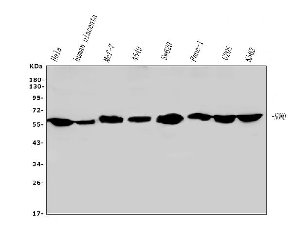
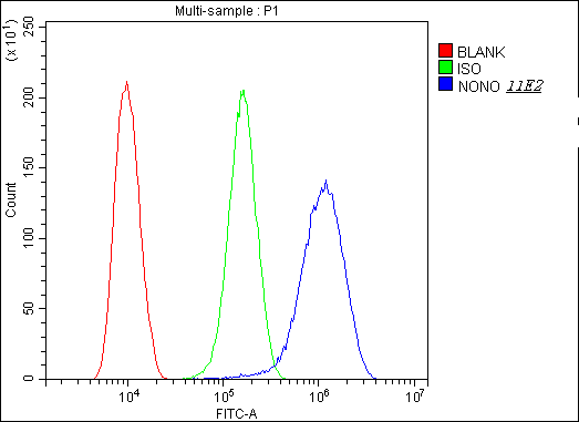
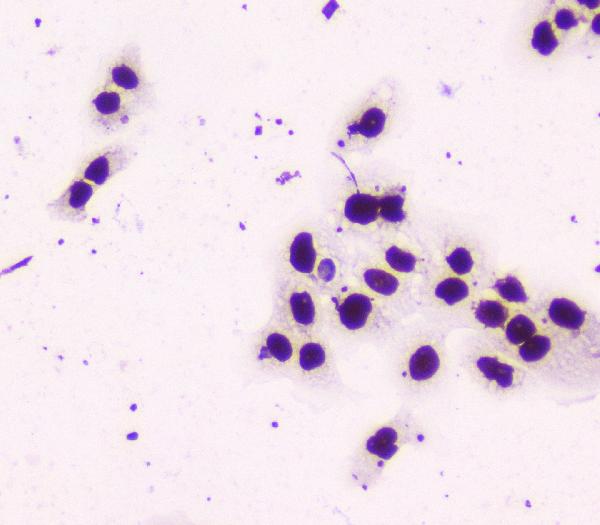
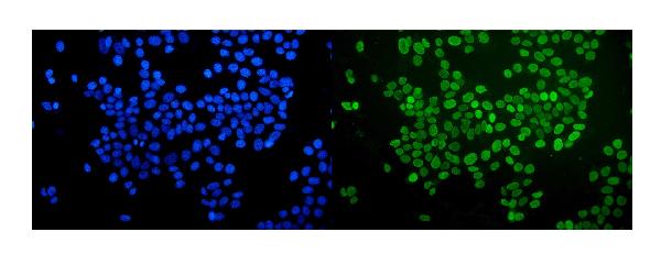
- 产品详情
- 实验流程
- 背景知识
Application 
| WB, IF, ICC, FC |
|---|---|
| Primary Accession | Q15233 |
| Host | Mouse |
| Isotype | Mouse IgG1 |
| Reactivity | Human |
| Clonality | Monoclonal |
| Format | Lyophilized |
| Description | Anti-nmt55 p54nrb NONO Antibody Picoband™ (monoclonal, 11E2) . Tested in Flow Cytometry, IF, ICC, WB applications. This antibody reacts with Human. |
| Gene ID | 4841 |
|---|---|
| Other Names | Non-POU domain-containing octamer-binding protein, NonO protein, 54 kDa nuclear RNA- and DNA-binding protein, p54(nrb), p54nrb, 55 kDa nuclear protein, NMT55, DNA-binding p52/p100 complex, 52 kDa subunit, NONO {ECO:0000303|PubMed:9393982, ECO:0000312|HGNC:HGNC:7871} |
| Calculated MW | 54232 Da |
| Application Details | Western blot, 0.1-0.5 µg/ml Immunocytochemistry, 0.5-1 µg/ml Immunocytochemistry/Immunofluorescence, 5 µg/ml Flow Cytometry, 1-3 µg/1x10^6 cells |
| Subcellular Localization | Nucleus. Nucleus, nucleolus. Nucleus speckle. Detected in punctate subnuclear structures often located adjacent to splicing speckles, called paraspeckles. |
| Tissue Specificity | Heart, brain, placenta, lung, liver, skeletal muscle, kidney and pancreas. Also found in a number of breast tumor cell lines. |
| Source | Eukaryota |
| Contents | Each vial contains 4mg Trehalose, 0.9mg NaCl, 0.2mg Na2HPO4, 0.05mg NaN3. |
| Clone Names | Clone: 11E2 |
| Immunogen | A synthetic peptide corresponding to a sequence at the N-terminus of human nmt55 p54nrb, identical to the related mouse and rat sequences. |
| Cross Reactivity | No cross-reactivity with other proteins. |
| Storage | Store at -20˚C for one year from date of receipt. After reconstitution, at 4˚C for one month. It can also be aliquotted and stored frozen at -20˚C for six months. Avoid repeated freeze-thaw cycles. |
For Research Use Only. Not For Use In Diagnostic Procedures.
| Name | NONO {ECO:0000303|PubMed:9393982, ECO:0000312|HGNC:HGNC:7871} |
|---|---|
| Function | DNA- and RNA binding protein, involved in several nuclear processes (PubMed:11525732, PubMed:12403470, PubMed:26571461). Binds the conventional octamer sequence in double-stranded DNA (PubMed:11525732, PubMed:12403470, PubMed:26571461). Also binds single- stranded DNA and RNA at a site independent of the duplex site (PubMed:11525732, PubMed:12403470, PubMed:26571461). Involved in pre- mRNA splicing, probably as a heterodimer with SFPQ (PubMed:11525732, PubMed:12403470, PubMed:26571461). Interacts with U5 snRNA, probably by binding to a purine-rich sequence located on the 3' side of U5 snRNA stem 1b (PubMed:12403470). Together with PSPC1, required for the formation of nuclear paraspeckles (PubMed:22416126). The SFPQ-NONO heteromer associated with MATR3 may play a role in nuclear retention of defective RNAs (PubMed:11525732). The SFPQ-NONO heteromer may be involved in DNA unwinding by modulating the function of topoisomerase I/TOP1 (PubMed:10858305). The SFPQ-NONO heteromer may be involved in DNA non-homologous end joining (NHEJ) required for double-strand break repair and V(D)J recombination and may stabilize paired DNA ends (PubMed:15590677). In vitro, the complex strongly stimulates DNA end joining, binds directly to the DNA substrates and cooperates with the Ku70/G22P1-Ku80/XRCC5 (Ku) dimer to establish a functional preligation complex (PubMed:15590677). NONO is involved in transcriptional regulation. The SFPQ-NONO-NR5A1 complex binds to the CYP17 promoter and regulates basal and cAMP-dependent transcriptional activity (PubMed:11897684). NONO binds to an enhancer element in long terminal repeats of endogenous intracisternal A particles (IAPs) and activates transcription (By similarity). Regulates the circadian clock by repressing the transcriptional activator activity of the CLOCK-BMAL1 heterodimer (By similarity). Important for the functional organization of GABAergic synapses (By similarity). Plays a specific and important role in the regulation of synaptic RNAs and GPHN/gephyrin scaffold structure, through the regulation of GABRA2 transcript (By similarity). Plays a key role during neuronal differentiation by recruiting TET1 to genomic loci and thereby regulating 5-hydroxymethylcytosine levels (By similarity). Plays a role in the regulation of DNA virus-mediated innate immune response by assembling into the HDP-RNP complex, a complex that serves as a platform for IRF3 phosphorylation and subsequent innate immune response activation through the cGAS-STING pathway (PubMed:28712728, PubMed:30270045). Promotes activation of the cGAS-STING pathway in response to HIV-2 infection: acts by interacting with HIV-2 Capsid protein p24, thereby promoting detection of viral DNA by CGAS, leading to CGAS-mediated inmmune activation (PubMed:30270045). In contrast, the weak interaction with HIV-1 Capsid protein p24 does not allow activation of the cGAS-STING pathway (PubMed:30270045). |
| Cellular Location | Nucleus. Nucleus, nucleolus. Nucleus speckle. Chromosome {ECO:0000250|UniProtKB:Q99K48}. Note=Detected in punctate subnuclear structures often located adjacent to splicing speckles, called paraspeckles. |
| Tissue Location | Heart, brain, placenta, lung, liver, skeletal muscle, kidney and pancreas. Also found in a number of breast tumor cell lines. |
Provided below are standard protocols that you may find useful for product applications.
BACKGROUND
Non-POU domain-containing octamer-binding protein is a protein that in humans is encoded by the NONO gene. This gene encodes an RNA-binding protein which plays various roles in the nucleus, including transcriptional regulation and RNA splicing. A rearrangement between this gene and the transcription factor E3 gene has been observed in papillary renal cell carcinoma. Alternatively spliced transcript variants have been described. Pseudogenes exist on Chromosomes 2 and 16.

终于等到您。ABCEPTA(百远生物)抗体产品。
点击下方“我要评价 ”按钮提交您的反馈信息,您的反馈和评价是我们最宝贵的财富之一,
我们将在1-3个工作日内处理您的反馈信息。
如有疑问,联系:0512-88856768 tech-china@abcepta.com.


