


 癌症的基本特征包括细胞增殖、血管生成、迁移、凋亡逃避机制和细胞永生等。找到癌症发生过程中这些通路的关键标记物和对应的抗体用于检测至关重要。
 为您推荐一个泛素化位点预测神器——泛素化分析工具,可以为您的蛋白的泛素化位点作出预测和评分。
 细胞自噬受体图形绘图工具为你的蛋白的细胞受体结合位点作出预测和评分,识别结合到自噬通路中的蛋白是非常重要的,便于让我们理解自噬在正常生理、病理过程中的作用,如发育、细胞分化、神经退化性疾病、压力条件下、感染和癌症。
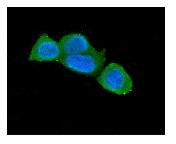
Anti-Caspase-3 Antibody Picoband™ (monoclonal, 15G8)
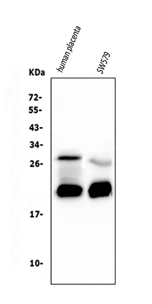
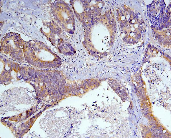
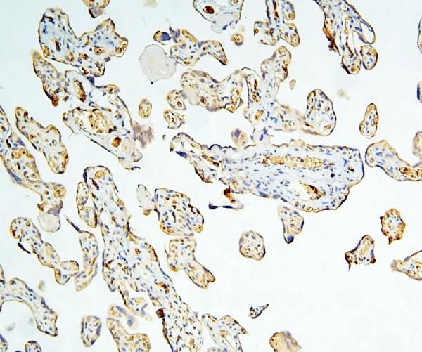
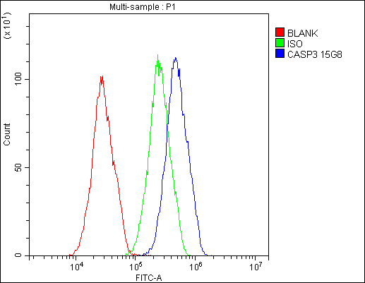
- 产品详情
- 实验流程
- 背景知识
Application 
| WB, IHC, IF, ICC, FC |
|---|---|
| Primary Accession | P42574 |
| Host | Mouse |
| Isotype | Mouse IgG1 |
| Reactivity | Human |
| Clonality | Monoclonal |
| Format | Lyophilized |
| Description | Anti-Caspase-3 Antibody Picoband™ (monoclonal, 15G8) . Tested in Flow Cytometry, IF, IHC, ICC, WB applications. This antibody reacts with Human. |
| Reconstitution | Add 0.2ml of distilled water will yield a concentration of 500 µg/ml. |
| Gene ID | 836 |
|---|---|
| Other Names | Caspase-3, CASP-3, 3.4.22.56, Apopain, Cysteine protease CPP32, CPP-32, Protein Yama, SREBP cleavage activity 1, SCA-1, Caspase-3 subunit p17, Caspase-3 subunit p12, CASP3, CPP32 {ECO:0000303|PubMed:7983002} |
| Calculated MW | 31608 Da |
| Application Details | Western blot, 0.1-0.5 µg/ml Immunohistochemistry (Paraffin-embedded Section), 0.5-1 µg/ml Immunocytochemistry/Immunofluorescence, 2 µg/ml, Human Flow Cytometry, 1-3 µg/1x10^6 cells |
| Subcellular Localization | Cytoplasm. |
| Tissue Specificity | Highly expressed in lung, spleen, heart, liver and kidney. Moderate levels in brain and skeletal muscle, and low in testis. Also found in many cell lines, highest expression in cells of the immune system. |
| Source | Eukaryota |
| Contents | Each vial contains 4mg Trehalose, 0.9mg NaCl, 0.2mg Na2HPO4, 0.05mg NaN3. |
| Clone Names | Clone: 15G8 |
| Immunogen | E.coli-derived human Caspase-3 recombinant protein (Position: T67-D175). Human Caspase-3 shares 86% and 90% amino acid (aa) sequence identity with mouse and rat Caspase-3, respectively. |
| Cross Reactivity | No cross-reactivity with other proteins. |
| Storage | Store at -20˚C for one year from date of receipt. After reconstitution, at 4˚C for one month. It can also be aliquotted and stored frozen at -20˚C for six months. Avoid repeated freeze-thaw cycles. |
For Research Use Only. Not For Use In Diagnostic Procedures.
| Name | CASP3 |
|---|---|
| Synonyms | CPP32 {ECO:0000303|PubMed:7983002} |
| Function | Thiol protease that acts as a major effector caspase involved in the execution phase of apoptosis (PubMed:18723680, PubMed:20566630, PubMed:23650375, PubMed:35338844, PubMed:35446120, PubMed:7596430). Following cleavage and activation by initiator caspases (CASP8, CASP9 and/or CASP10), mediates execution of apoptosis by catalyzing cleavage of many proteins (PubMed:18723680, PubMed:20566630, PubMed:23650375, PubMed:7596430). At the onset of apoptosis, it proteolytically cleaves poly(ADP-ribose) polymerase PARP1 at a '216-Asp-|-Gly-217' bond (PubMed:10497198, PubMed:16374543, PubMed:7596430, PubMed:7774019). Cleaves and activates sterol regulatory element binding proteins (SREBPs) between the basic helix-loop-helix leucine zipper domain and the membrane attachment domain (By similarity). Cleaves and activates caspase-6, -7 and -9 (CASP6, CASP7 and CASP9, respectively) (PubMed:7596430). Cleaves and inactivates interleukin-18 (IL18) (PubMed:37993714, PubMed:9334240). Involved in the cleavage of huntingtin (PubMed:8696339). Triggers cell adhesion in sympathetic neurons through RET cleavage (PubMed:21357690). Also involved in axon pruning during brain development (By similarity). Cleaves DSG2 in response to apoptosis resulting in a loss of full length DSG2 at desmosome cell junctions and subsequent loss of cell-cell adhesion (PubMed:17559062). Also cleaves JUP in response to apoptosis (PubMed:17559062). Cleaves and inhibits serine/threonine-protein kinase AKT1 in response to oxidative stress (PubMed:23152800). Acts as an inhibitor of type I interferon production during virus-induced apoptosis by mediating cleavage of antiviral proteins CGAS, IRF3 and MAVS, thereby preventing cytokine overproduction (PubMed:30878284). Also involved in pyroptosis by mediating cleavage and activation of gasdermin-E (GSDME) (PubMed:35338844, PubMed:35446120). Cleaves XRCC4 and phospholipid scramblase proteins XKR4, XKR8 and XKR9, leading to promote phosphatidylserine exposure on apoptotic cell surface (PubMed:23845944, PubMed:33725486). Cleaves BIRC6 following inhibition of BIRC6-caspase binding by DIABLO/SMAC (PubMed:36758104, PubMed:36758106). |
| Cellular Location | Cytoplasm. |
| Tissue Location | Highly expressed in lung, spleen, heart, liver and kidney. Moderate levels in brain and skeletal muscle, and low in testis. Also found in many cell lines, highest expression in cells of the immune system. |
Provided below are standard protocols that you may find useful for product applications.
BACKGROUND
Caspase 3 is a caspase protein which interacts with Survivin, XIAP, CFLAR, Caspase 8, HCLS1, Deleted in Colorectal Cancer, TRAF3 and GroEL. This gene which is located on 4q35 encodes a protein that is a member of the cysteine-aspartic acid protease (caspase) family. Sequential activation of caspases plays a central role in the execution-phase of cell apoptosis. Caspases exist as inactive proenzymes that undergo proteolytic processing at conserved aspartic residues to produce two subunits, large and small, that dimerize to form the active enzyme. It is the predominant caspase involved in the cleavage of amyloid-beta 4A precursor protein, which is associated with neuronal death in Alzheimer's disease. And the caspase-3 activation in heart failure sequentially cleaves SRF and generates a truncated SRF that appears to function as a dominant-negative transcription factor. Additionally, the caspase-3 influence on bone mineral density should be considered in any in vivo application of caspase-3 inhibitors to the treatment of human disease. In erythroid precursors undergoing terminal differentiation, Hsp70 prevents active CASP3 from cleaving GATA1 and inducing apoptosis.

终于等到您。ABCEPTA(百远生物)抗体产品。
点击下方“我要评价 ”按钮提交您的反馈信息,您的反馈和评价是我们最宝贵的财富之一,
我们将在1-3个工作日内处理您的反馈信息。
如有疑问,联系:0512-88856768 tech-china@abcepta.com.


