


 癌症的基本特征包括细胞增殖、血管生成、迁移、凋亡逃避机制和细胞永生等。找到癌症发生过程中这些通路的关键标记物和对应的抗体用于检测至关重要。
 为您推荐一个泛素化位点预测神器——泛素化分析工具,可以为您的蛋白的泛素化位点作出预测和评分。
 细胞自噬受体图形绘图工具为你的蛋白的细胞受体结合位点作出预测和评分,识别结合到自噬通路中的蛋白是非常重要的,便于让我们理解自噬在正常生理、病理过程中的作用,如发育、细胞分化、神经退化性疾病、压力条件下、感染和癌症。
Anti-HAUSP/USP7 Antibody Picoband™ (monoclonal, 5E2)
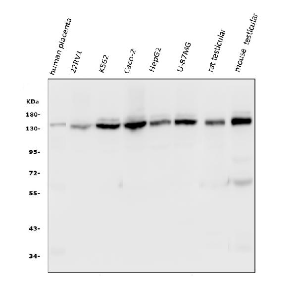
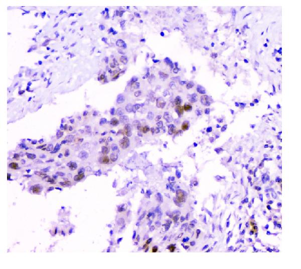
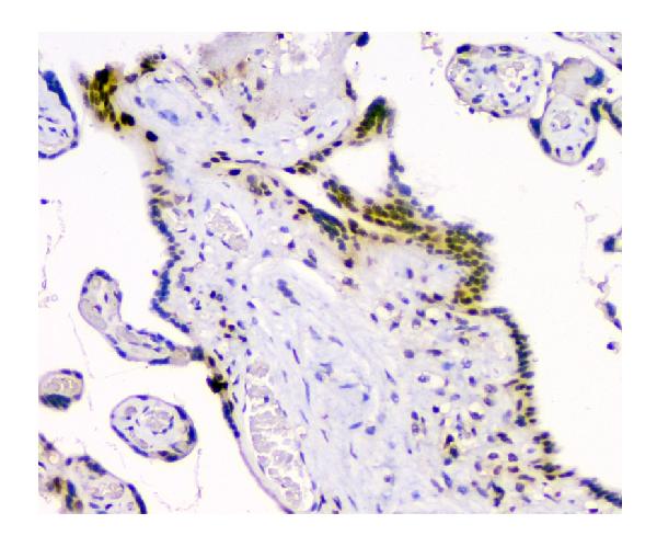
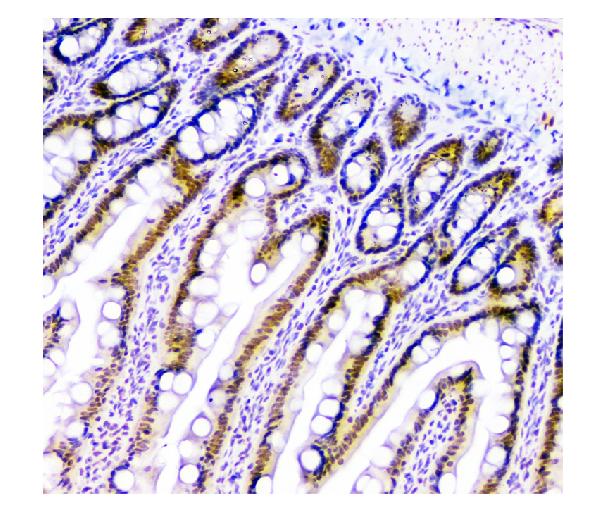
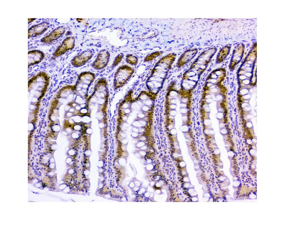
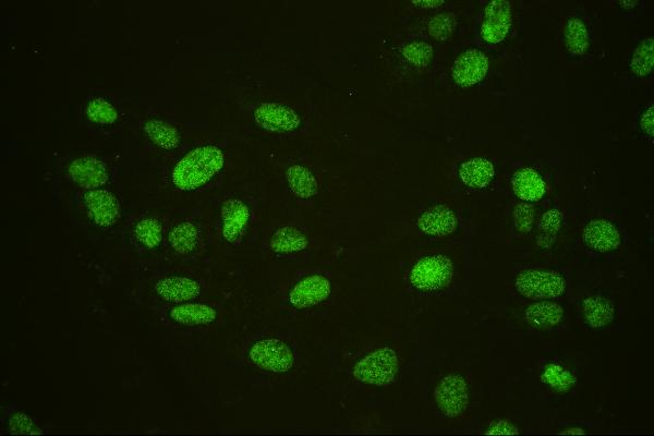
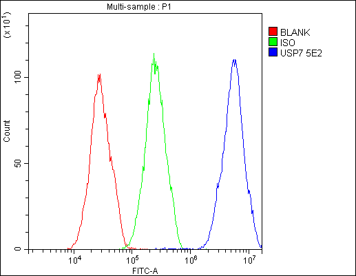
- 产品详情
- 实验流程
- 背景知识
Application 
| WB, IHC, IF, ICC, FC |
|---|---|
| Primary Accession | Q93009 |
| Host | Mouse |
| Isotype | Mouse IgG2b |
| Reactivity | Rat, Human, Mouse |
| Clonality | Monoclonal |
| Format | Lyophilized |
| Description | Anti-HAUSP/USP7 Antibody Picoband™ (monoclonal, 5E2) . Tested in Flow Cytometry, IF, IHC, ICC, WB applications. This antibody reacts with Human, Mouse, Rat. |
| Reconstitution | Add 0.2ml of distilled water will yield a concentration of 500 µg/ml. |
| Gene ID | 7874 |
|---|---|
| Other Names | Ubiquitin carboxyl-terminal hydrolase 7, 3.4.19.12, Deubiquitinating enzyme 7, Herpesvirus-associated ubiquitin-specific protease, Ubiquitin thioesterase 7, Ubiquitin-specific-processing protease 7, USP7 {ECO:0000303|PubMed:12093161, ECO:0000312|HGNC:HGNC:12630} |
| Calculated MW | 128302 Da |
| Application Details | Western blot, 0.1-0.5 µg/ml Immunohistochemistry (Paraffin-embedded Section), 0.5-1 µg/ml Immunocytochemistry/Immunofluorescence, 2 µg/ml Flow Cytometry, 1-3 µg/1x10^6 cells |
| Subcellular Localization | Nucleus. PML body. Cytoplasm. Chromosome. |
| Tissue Specificity | Widely expressed. Overexpressed in prostate cancer. |
| Source | Eukaryota |
| Contents | Each vial contains 4mg Trehalose, 0.9mg NaCl, 0.2mg Na2HPO4, 0.05mg NaN3. |
| Clone Names | Clone: Clone: 5E2 |
| Immunogen | E.coli-derived human HAUSP/USP7 recombinant protein (Position: L258-D483). |
| Cross Reactivity | No cross-reactivity with other proteins. |
| Storage | Store at -20˚C for one year from date of receipt. After reconstitution, at 4˚C for one month. It can also be aliquotted and stored frozen at -20˚C for six months. Avoid repeated freeze-thaw cycles. |
For Research Use Only. Not For Use In Diagnostic Procedures.
| Name | USP7 {ECO:0000303|PubMed:12093161, ECO:0000312|HGNC:HGNC:12630} |
|---|---|
| Function | Hydrolase that deubiquitinates target proteins such as ARMC5, FOXO4, DEPTOR, KAT5, p53/TP53, MDM2, ERCC6, DNMT1, UHRF1, PTEN, KMT2E/MLL5 and DAXX (PubMed:11923872, PubMed:15053880, PubMed:16964248, PubMed:18716620, PubMed:25283148, PubMed:25865756, PubMed:26678539, PubMed:28655758, PubMed:33544460, PubMed:35216969). Together with DAXX, prevents MDM2 self-ubiquitination and enhances the E3 ligase activity of MDM2 towards p53/TP53, thereby promoting p53/TP53 ubiquitination and proteasomal degradation (PubMed:15053880, PubMed:16845383, PubMed:18566590, PubMed:20153724). Deubiquitinates p53/TP53, preventing degradation of p53/TP53, and enhances p53/TP53-dependent transcription regulation, cell growth repression and apoptosis (PubMed:25283148). Deubiquitinates p53/TP53 and MDM2 and strongly stabilizes p53/TP53 even in the presence of excess MDM2, and also induces p53/TP53-dependent cell growth repression and apoptosis (PubMed:11923872, PubMed:26786098). Deubiquitination of FOXO4 in presence of hydrogen peroxide is not dependent on p53/TP53 and inhibits FOXO4-induced transcriptional activity (PubMed:16964248). In association with DAXX, is involved in the deubiquitination and translocation of PTEN from the nucleus to the cytoplasm, both processes that are counteracted by PML (PubMed:18716620). Deubiquitinates KMT2E/MLL5 preventing KMT2E/MLL5 proteasomal-mediated degradation (PubMed:26678539). Involved in cell proliferation during early embryonic development. Involved in transcription-coupled nucleotide excision repair (TC-NER) in response to UV damage: recruited to DNA damage sites following interaction with KIAA1530/UVSSA and promotes deubiquitination of ERCC6, preventing UV- induced degradation of ERCC6 (PubMed:22466611, PubMed:22466612). Involved in maintenance of DNA methylation via its interaction with UHRF1 and DNMT1: acts by mediating deubiquitination of UHRF1 and DNMT1, preventing their degradation and promoting DNA methylation by DNMT1 (PubMed:21745816, PubMed:22411829). Deubiquitinates alkylation repair enzyme ALKBH3. OTUD4 recruits USP7 and USP9X to stabilize ALKBH3, thereby promoting the repair of alkylated DNA lesions (PubMed:25944111). Acts as a chromatin regulator via its association with the Polycomb group (PcG) multiprotein PRC1-like complex; may act by deubiquitinating components of the PRC1-like complex (PubMed:20601937). Able to mediate deubiquitination of histone H2B; it is however unsure whether this activity takes place in vivo (PubMed:20601937). Exhibits a preference towards 'Lys-48'-linked ubiquitin chains (PubMed:22689415). Increases regulatory T-cells (Treg) suppressive capacity by deubiquitinating and stabilizing the transcription factor FOXP3 which is crucial for Treg cell function (PubMed:23973222). Plays a role in the maintenance of the circadian clock periodicity via deubiquitination and stabilization of the CRY1 and CRY2 proteins (PubMed:27123980). Deubiquitinates REST, thereby stabilizing REST and promoting the maintenance of neural progenitor cells (PubMed:21258371). Deubiquitinates SIRT7, inhibiting SIRT7 histone deacetylase activity and regulating gluconeogenesis (PubMed:28655758). Involved in the regulation of WASH-dependent actin polymerization at the surface of endosomes and the regulation of endosomal protein recycling (PubMed:26365382). It maintains optimal WASH complex activity and precise F-actin levels via deubiquitination of TRIM27 and WASHC1 (PubMed:26365382). Mediates the deubiquitination of phosphorylated DEPTOR, promoting its stability and leading to decreased mTORC1 signaling (PubMed:35216969). Deubiquitinates H2AB1, H2BC4 and H2BC22 (By similarity). |
| Cellular Location | Nucleus. Cytoplasm Nucleus, PML body. Chromosome. Note=Present in a minority of ND10 nuclear bodies (PubMed:9034339). Association with ICP0/VMW110 at early times of infection leads to an increased proportion of USP7-containing ND10 (PubMed:9034339). Colocalizes with ATXN1 in the nucleus (PubMed:12093161). Colocalized with DAXX in speckled structures (PubMed:16845383). Colocalized with PML and PTEN in promyelocytic leukemia protein (PML) nuclear bodies (PubMed:18716620). Interaction with FAM170A promotes its nuclear translocation (By similarity) {ECO:0000250|UniProtKB:Q6A4J8, ECO:0000269|PubMed:12093161, ECO:0000269|PubMed:16845383, ECO:0000269|PubMed:18716620, ECO:0000269|PubMed:9034339} |
| Tissue Location | Expressed in neural progenitor cells (at protein level) (PubMed:21258371). Widely expressed. Overexpressed in prostate cancer. |
Provided below are standard protocols that you may find useful for product applications.
BACKGROUND
Ubiquitin-specific-processing protease 7 (USP7), also known as ubiquitin carboxyl-terminal hydrolase 7 or herpesvirus-associated ubiquitin-specific protease (HAUSP), is an enzyme that in humans is encoded by the USP7 gene. The protein encoded by this gene belongs to the peptidase C19 family, which includes ubiquitinyl hydrolases. This protein deubiquitinates target proteins such as p53 (a tumor suppressor protein) and WASH (essential for endosomal protein recycling), and regulates their activities by counteracting the opposing ubiquitin ligase activity of proteins such as HDM2 and TRIM27, involved in the respective process. Mutations in this gene have been implicated in a neurodevelopmental disorder.

终于等到您。ABCEPTA(百远生物)抗体产品。
点击下方“我要评价 ”按钮提交您的反馈信息,您的反馈和评价是我们最宝贵的财富之一,
我们将在1-3个工作日内处理您的反馈信息。
如有疑问,联系:0512-88856768 tech-china@abcepta.com.


