


 癌症的基本特征包括细胞增殖、血管生成、迁移、凋亡逃避机制和细胞永生等。找到癌症发生过程中这些通路的关键标记物和对应的抗体用于检测至关重要。
 为您推荐一个泛素化位点预测神器——泛素化分析工具,可以为您的蛋白的泛素化位点作出预测和评分。
 细胞自噬受体图形绘图工具为你的蛋白的细胞受体结合位点作出预测和评分,识别结合到自噬通路中的蛋白是非常重要的,便于让我们理解自噬在正常生理、病理过程中的作用,如发育、细胞分化、神经退化性疾病、压力条件下、感染和癌症。
Anti-RBPJK/RBPJ Picoband™ Antibody (monoclonal, 6G4)
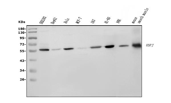
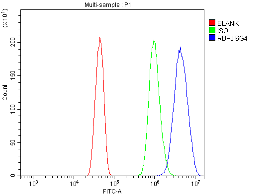
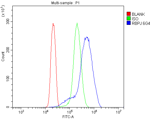
- 产品详情
- 实验流程
- 背景知识
Application 
| WB, FC |
|---|---|
| Primary Accession | Q06330 |
| Host | Mouse |
| Isotype | Mouse IgG2b |
| Reactivity | Rat, Human, Mouse |
| Clonality | Monoclonal |
| Format | Lyophilized |
| Description | Anti-RBPJK/RBPJ Picoband™ Antibody (monoclonal, 6G4) . Tested in Flow Cytometry, WB applications. This antibody reacts with Human, Mouse, Rat. |
| Reconstitution | Add 0.2ml of distilled water will yield a concentration of 500ug/ml. |
| Gene ID | 3516 |
|---|---|
| Other Names | Recombining binding protein suppressor of hairless, CBF-1, J kappa-recombination signal-binding protein, RBP-J kappa, RBP-J, RBP-JK, Renal carcinoma antigen NY-REN-30, RBPJ (HGNC:5724) |
| Calculated MW | 55637 Da |
| Application Details | Western blot, 0.25-0.5 µg/ml, Human, Mouse, Rat Flow Cytometry, 1-3 µg/1x10^6 cells, Human, Mouse |
| Source | Eukaryota |
| Contents | Each vial contains 4mg Trehalose, 0.9mg NaCl and 0.2mg Na2HPO4. |
| Clone Names | Clone: 6G4 |
| Immunogen | E.coli-derived human RBPJK/RBPJ recombinant protein (Position: K41-Q467). |
| Purification | Immunogen affinity purified. |
| Storage | Store at -20˚C for one year from date of receipt. After reconstitution, at 4˚C for one month. It can also be aliquotted and stored frozen at -20˚C for six months. Avoid repeated freeze-thaw cycles. |
For Research Use Only. Not For Use In Diagnostic Procedures.
| Name | RBPJ (HGNC:5724) |
|---|---|
| Function | Transcriptional regulator that plays a central role in Notch signaling, a signaling pathway involved in cell-cell communication that regulates a broad spectrum of cell-fate determinations (PubMed:41086914, PubMed:36129980). Acts as a transcriptional repressor when it is not associated with Notch proteins. When associated with some NICD product of Notch proteins (Notch intracellular domain), it acts as a transcriptional activator that activates transcription of Notch target genes. Probably represses or activates transcription via the recruitment of chromatin remodeling complexes containing histone deacetylase or histone acetylase proteins, respectively. Specifically binds to the immunoglobulin kappa-type J segment recombination signal sequence. Binds specifically to methylated DNA (PubMed:21991380). Binds to the oxygen responsive element of COX4I2 and activates its transcription under hypoxia conditions (4% oxygen) (PubMed:23303788). Negatively regulates the phagocyte oxidative burst in response to bacterial infection by repressing transcription of NADPH oxidase subunits (By similarity). |
| Cellular Location | Nucleus. Cytoplasm. Note=Mainly nuclear, upon interaction with RITA/C12orf52, translocates to the cytoplasm, down- regulating the Notch signaling pathway |
Provided below are standard protocols that you may find useful for product applications.
BACKGROUND
Recombination signal binding protein for immunoglobulin kappa J region is a protein that in humans is encoded by the RBPJ gene. It is mapped to 4p15.2. The protein encoded by this gene is a transcriptional regulator important in the Notch signaling pathway. The encoded protein acts as a repressor when not bound to Notch proteins and an activator when bound to Notch proteins. It is thought to function by recruiting chromatin remodeling complexes containing histone deacetylase or histone acetylase proteins to Notch signaling pathway genes. Several transcript variants encoding different isoforms have been found for this gene, and several pseudogenes of this gene exist on chromosome 9.

终于等到您。ABCEPTA(百远生物)抗体产品。
点击下方“我要评价 ”按钮提交您的反馈信息,您的反馈和评价是我们最宝贵的财富之一,
我们将在1-3个工作日内处理您的反馈信息。
如有疑问,联系:0512-88856768 tech-china@abcepta.com.


