


 癌症的基本特征包括细胞增殖、血管生成、迁移、凋亡逃避机制和细胞永生等。找到癌症发生过程中这些通路的关键标记物和对应的抗体用于检测至关重要。
 为您推荐一个泛素化位点预测神器——泛素化分析工具,可以为您的蛋白的泛素化位点作出预测和评分。
 细胞自噬受体图形绘图工具为你的蛋白的细胞受体结合位点作出预测和评分,识别结合到自噬通路中的蛋白是非常重要的,便于让我们理解自噬在正常生理、病理过程中的作用,如发育、细胞分化、神经退化性疾病、压力条件下、感染和癌症。
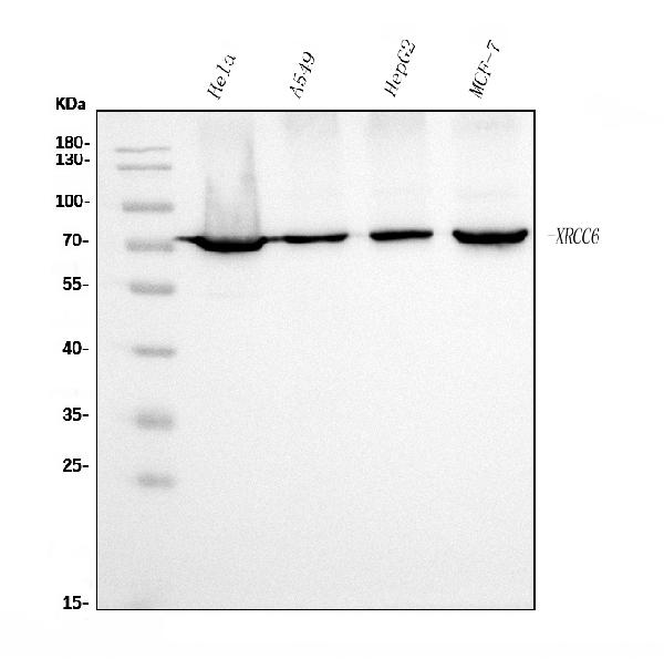
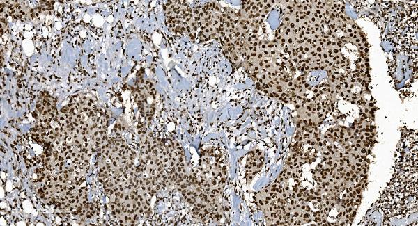
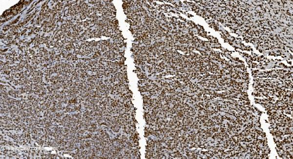
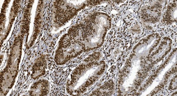
Anti-Ku70 Antibody Picoband™ (monoclonal, 9B6)
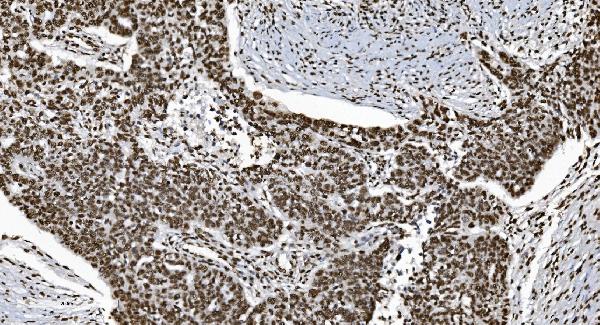
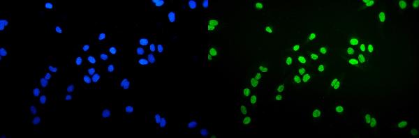
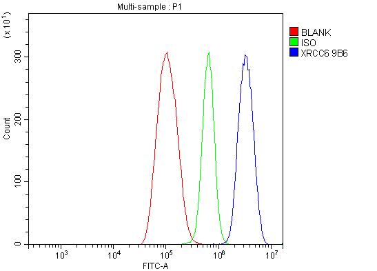
- 产品详情
- 实验流程
- 背景知识
Application 
| WB, IHC, IF, ICC, FC |
|---|---|
| Primary Accession | P12956 |
| Host | Mouse |
| Isotype | Mouse IgG2b |
| Reactivity | Human |
| Clonality | Monoclonal |
| Format | Lyophilized |
| Description | Anti-Ku70 Antibody Picoband™ (monoclonal, 9B6) . Tested in Flow Cytometry, IF, IHC, ICC, WB applications. This antibody reacts with Human. |
| Reconstitution | Add 0.2ml of distilled water will yield a concentration of 500ug/ml. |
| Gene ID | 2547 |
|---|---|
| Other Names | X-ray repair cross-complementing protein 6, 3.6.4.-, 4.2.99.-, 5'-deoxyribose-5-phosphate lyase Ku70, 5'-dRP lyase Ku70, 70 kDa subunit of Ku antigen, ATP-dependent DNA helicase 2 subunit 1, ATP-dependent DNA helicase II 70 kDa subunit, CTC box-binding factor 75 kDa subunit, CTC75, CTCBF, DNA repair protein XRCC6, Lupus Ku autoantigen protein p70, Ku70, Thyroid-lupus autoantigen, TLAA, X-ray repair complementing defective repair in Chinese hamster cells 6, XRCC6, G22P1 |
| Calculated MW | 69843 Da |
| Application Details | Western blot, 0.25-0.5 µg/ml, Human Immunohistochemistry (Paraffin-embedded Section), 2-5 µg/ml, Human Immunocytochemistry/Immunofluorescence, 5 µg/ml, Human Flow Cytometry, 1-3 µg/1x10^6 cells, Human |
| Source | Eukaryota |
| Contents | Each vial contains 4mg Trehalose, 0.9mg NaCl and 0.2mg Na2HPO4. |
| Clone Names | Clone: 9B6 |
| Immunogen | A synthetic peptide corresponding to a sequence at C-terminus of human Ku70, different from the related mouse sequence by one amino acid. |
| Purification | Immunogen affinity purified. |
| Storage | Store at -20˚C for one year from date of receipt. After reconstitution, at 4˚C for one month. It can also be aliquotted and stored frozen at -20˚C for six months. Avoid repeated freeze-thaw cycles. |
For Research Use Only. Not For Use In Diagnostic Procedures.
| Name | XRCC6 (HGNC:4055) |
|---|---|
| Synonyms | G22P1 |
| Function | DNA-binding protein critical for the DNA damage response, specifically in repairing double-strand breaks (DSBs) via the classical non-homologous end joining (NHEJ) pathway. It forms a heterodimer with XRCC5 (Ku80), creating the Ku70:Ku80 heterodimer (Ku complex), which serves as a DNA end-binding complex. It primarily binds DSBs and recruits essential repair factors, assembling the core long-range NHEJ complex to facilitate the alignment and ligation of broken DNA ends (PubMed:11493912, PubMed:20493174, PubMed:33854234, PubMed:34352203, PubMed:9742108). This pathway ensures the rapid repair of cytotoxic and mutagenic DSBs and contributes to the generation of diversity in T-cell receptors and antibodies through mechanisms such as V(D)J recombination (PubMed:9742108). Likely acts as a 5'-deoxyribose-5-phosphate lyase (5'-dRP lyase), catalyzing the beta-elimination of the 5'-deoxyribose- 5-phosphate at abasic sites near DSBs. This activity cleans the termini of abasic sites, a common form of nucleotide damage, preparing broken ends for ligation (PubMed:20383123). It may also possess 3'-5' DNA helicase activity, although this has not been confirmed in vivo, and its physiological significance remains unclear (PubMed:7957065). Beyond DNA repair, the protein contributes to telomere maintenance (PubMed:29490055). It is also implicated in transcriptional regulation, acting as a cofactor for various transcription factors (PubMed:12145306, PubMed:8621488). It plays a role in the regulation of DNA virus-mediated innate immune response by assembling into the HDP- RNP complex, a complex that serves as a platform for IRF3 phosphorylation and subsequent innate immune response activation through the cGAS-STING pathway (PubMed:28712728). Can also bind RNAs and recruits PRKDC to a wide range of cellular RNAs, including the U3 small nucleolar RNA, playing a role in the biogenesis of ribosomal RNAs (PubMed:32103174). Additionally, it negatively regulates apoptosis by interacting with BAX, sequestering it from the mitochondria, and may possess deubiquitination activity targeting BAX (PubMed:15023334, PubMed:18362350, PubMed:35545041). |
| Cellular Location | Nucleus. Chromosome. Cytoplasm. Note=When trimethylated, localizes in the cytoplasm. |
Provided below are standard protocols that you may find useful for product applications.
BACKGROUND
XRCC6 (X-Ray Repair, Complementing Defective, In Chinese Hamster, 6), also called Ku70, G22P1 or TLAA, is a protein that in humans, is encoded by the XRCC6 gene. In addition, the XRCC6 gene encodes subunit p70 of the p70/p80 autoantigen which consists of 2 proteins of molecular mass of approximately 70,000 and 80,000 daltons that dimerize to form a 10 S DNA-binding complex. The XRCC6 gene is mapped to 22q13.2. XRCC6 and Mre11 are differentially expressed during meiosis. XRCC6 interacts with Baxa, a mediator of mitochondrial-dependent apoptosis. Disruption of both FANCC and XRCC6 suppressed sensitivity to crosslinking agents, diminished chromosome breaks, and reversed defective homologous recombination. Ku70 binds directly to free DNA ends, committing them to NHEJ repair. In early meiotic prophase, however, when meiotic recombination is most probably initiated, Mre11 was abundant, whereas XRCC6 was not detectable.

终于等到您。ABCEPTA(百远生物)抗体产品。
点击下方“我要评价 ”按钮提交您的反馈信息,您的反馈和评价是我们最宝贵的财富之一,
我们将在1-3个工作日内处理您的反馈信息。
如有疑问,联系:0512-88856768 tech-china@abcepta.com.


