


 癌症的基本特征包括细胞增殖、血管生成、迁移、凋亡逃避机制和细胞永生等。找到癌症发生过程中这些通路的关键标记物和对应的抗体用于检测至关重要。
 为您推荐一个泛素化位点预测神器——泛素化分析工具,可以为您的蛋白的泛素化位点作出预测和评分。
 细胞自噬受体图形绘图工具为你的蛋白的细胞受体结合位点作出预测和评分,识别结合到自噬通路中的蛋白是非常重要的,便于让我们理解自噬在正常生理、病理过程中的作用,如发育、细胞分化、神经退化性疾病、压力条件下、感染和癌症。
Anti-INPPL1 Antibody Picoband™ (monoclonal, 8C13)
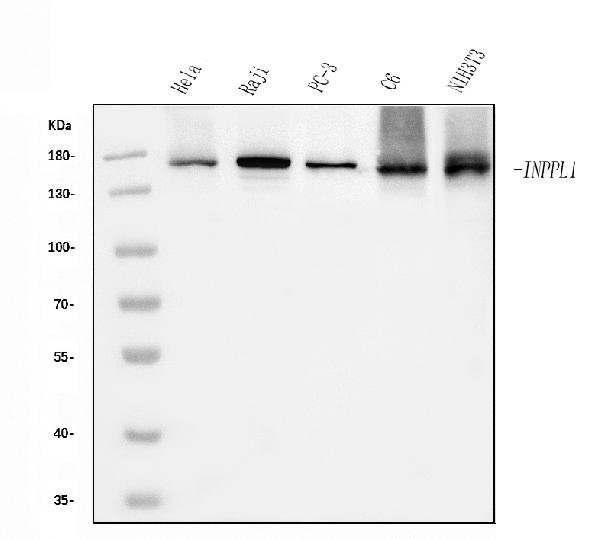
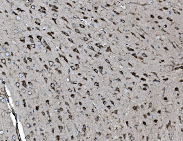
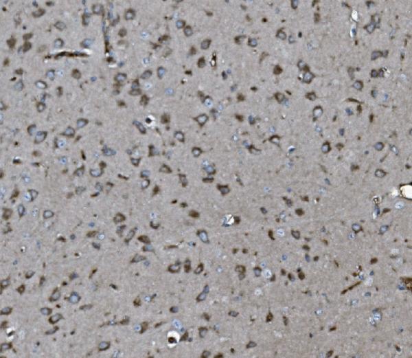
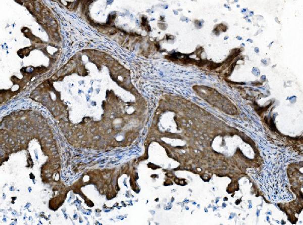
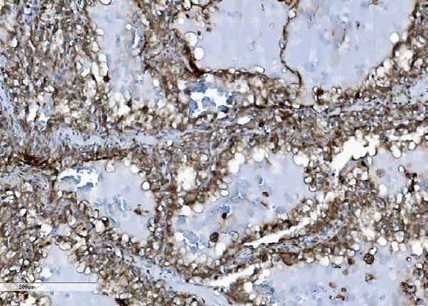
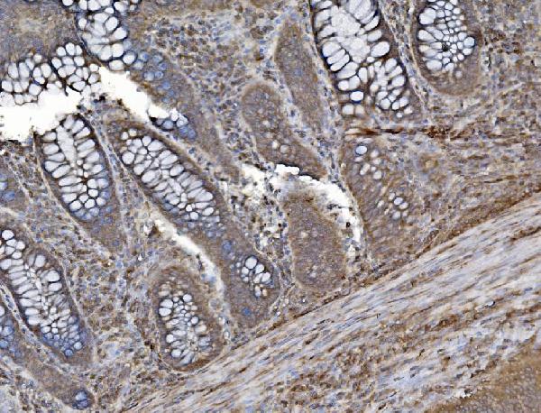
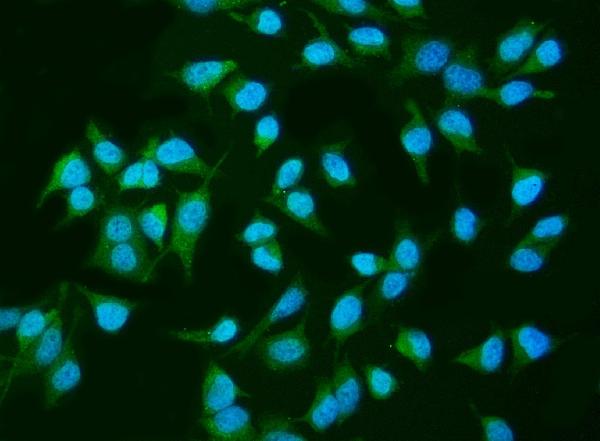
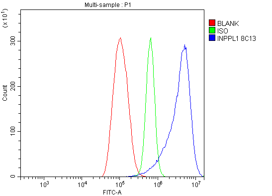
- 产品详情
- 实验流程
- 背景知识
Application 
| WB, IHC, IF, ICC, FC |
|---|---|
| Primary Accession | O15357 |
| Host | Mouse |
| Isotype | Mouse IgG2a |
| Reactivity | Rat, Human, Mouse |
| Clonality | Monoclonal |
| Format | Lyophilized |
| Description | Anti-INPPL1 Antibody Picoband™ (monoclonal, 8C13) . Tested in Flow Cytometry, IF, IHC, ICC, WB applications. This antibody reacts with Human, Mouse, Rat. |
| Reconstitution | Add 0.2ml of distilled water will yield a concentration of 500ug/ml. |
| Gene ID | 3636 |
|---|---|
| Other Names | Phosphatidylinositol 3, 4, 5-trisphosphate 5-phosphatase 2, 3.1.3.86, Inositol polyphosphate phosphatase-like protein 1, INPPL-1, Protein 51C, SH2 domain-containing inositol 5'-phosphatase 2 {ECO:0000312|HGNC:HGNC:6080}, SH2 domain-containing inositol phosphatase 2, SHIP-2, INPPL1 (HGNC:6080) |
| Calculated MW | 138599 Da |
| Application Details | Western blot, 0.25-0.5 µg/ml, Human, Mouse, Rat Immunohistochemistry (Paraffin-embedded Section), 2-5 µg/ml, Human, Mouse, Rat Immunocytochemistry/Immunofluorescence, 5 µg/ml, Human Flow Cytometry, 1-3 µg/1x10^6 cells, Human |
| Source | Eukaryota |
| Contents | Each vial contains 4mg Trehalose, 0.9mg NaCl and 0.2mg Na2HPO4. |
| Clone Names | Clone: 8C13 |
| Immunogen | E. coli-derived human INPPL1 recombinant protein (Position: R1172-K1258). |
| Purification | Immunogen affinity purified. |
| Storage | Store at -20˚C for one year from date of receipt. After reconstitution, at 4˚C for one month. It can also be aliquotted and stored frozen at -20˚C for six months. Avoid repeated freeze-thaw cycles. |
For Research Use Only. Not For Use In Diagnostic Procedures.
| Name | INPPL1 (HGNC:6080) |
|---|---|
| Function | Phosphatidylinositol (PtdIns) phosphatase that specifically hydrolyzes the 5-phosphate of phosphatidylinositol-3,4,5-trisphosphate (PtdIns(3,4,5)P3) to produce PtdIns(3,4)P2, thereby negatively regulating the PI3K (phosphoinositide 3-kinase) pathways (PubMed:16824732). Required for correct mitotic spindle orientation and therefore progression of mitosis (By similarity). Plays a central role in regulation of PI3K-dependent insulin signaling, although the precise molecular mechanisms and signaling pathways remain unclear (PubMed:9660833). While overexpression reduces both insulin-stimulated MAP kinase and Akt activation, its absence does not affect insulin signaling or GLUT4 trafficking (By similarity). Confers resistance to dietary obesity (By similarity). May act by regulating AKT2, but not AKT1, phosphorylation at the plasma membrane (By similarity). Part of a signaling pathway that regulates actin cytoskeleton remodeling (PubMed:11739414, PubMed:12676785). Required for the maintenance and dynamic remodeling of actin structures as well as in endocytosis, having a major impact on ligand-induced EGFR internalization and degradation (PubMed:15668240). Participates in regulation of cortical and submembraneous actin by hydrolyzing PtdIns(3,4,5)P3 thereby regulating membrane ruffling (PubMed:21624956). Regulates cell adhesion and cell spreading (PubMed:12235291). Required for HGF-mediated lamellipodium formation, cell scattering and spreading (PubMed:15735664). Acts as a negative regulator of EPHA2 receptor endocytosis by inhibiting via PI3K-dependent Rac1 activation (PubMed:17135240). Acts as a regulator of neuritogenesis by regulating PtdIns(3,4,5)P3 level and is required to form an initial protrusive pattern, and later, maintain proper neurite outgrowth (By similarity). Acts as a negative regulator of the FC-gamma-RIIA receptor (FCGR2A) (PubMed:12690104). Mediates signaling from the FC-gamma-RIIB receptor (FCGR2B), playing a central role in terminating signal transduction from activating immune/hematopoietic cell receptor systems (PubMed:11016922). Involved in EGF signaling pathway (PubMed:11349134). Upon stimulation by EGF, it is recruited by EGFR and dephosphorylates PtdIns(3,4,5)P3 (PubMed:11349134). Plays a negative role in regulating the PI3K-PKB pathway, possibly by inhibiting PKB activity (PubMed:11349134). Down-regulates Fc-gamma-R-mediated phagocytosis in macrophages independently of INPP5D/SHIP1 (By similarity). In macrophages, down-regulates NF-kappa-B-dependent gene transcription by regulating macrophage colony-stimulating factor (M-CSF)-induced signaling (By similarity). Plays a role in the localization of AURKA and NEDD9/HEF1 to the basolateral membrane at interphase in polarized cysts, thereby mediates cell cycle homeostasis, cell polarization and cilia assembly (By similarity). Additionally promotion of cilia growth is also facilitated by hydrolysis of (PtdIns(3,4,5)P3) to PtdIns(3,4)P2 (By similarity). Promotes formation of apical membrane-initiation sites during the initial stages of lumen formation via Rho family-induced actin filament organization and CTNNB1 localization to cell-cell contacts (By similarity). May also hydrolyze PtdIns(1,3,4,5)P4, and could thus affect the levels of the higher inositol polyphosphates like InsP6. Involved in endochondral ossification (PubMed:23273569). |
| Cellular Location | Cytoplasm, cytosol. Cytoplasm, cytoskeleton. Membrane; Peripheral membrane protein. Cell projection, filopodium. Cell projection, lamellipodium. Basal cell membrane {ECO:0000250|UniProtKB:F1PNY0}. Nucleus {ECO:0000250|UniProtKB:D7PF45} Nucleus speckle {ECO:0000250|UniProtKB:D7PF45}. Cytoplasm, cytoskeleton, spindle pole {ECO:0000250|UniProtKB:F1PNY0} Note=Translocates to membrane ruffles when activated, translocation is probably due to different mechanisms depending on the stimulus and cell type (PubMed:11739414). Partly translocated via its SH2 domain which mediates interaction with tyrosine phosphorylated receptors such as the FC-gamma-RIIB receptor (FCGR2B). Tyrosine phosphorylation may also participate in membrane localization. Insulin specifically stimulates its redistribution from the cytosol to the plasma membrane. Recruited to the membrane following M-CSF stimulation. In activated spreading platelets, localizes with actin at filopodia, lamellipodia and the central actin ring. |
| Tissue Location | Widely expressed, most prominently in skeletal muscle, heart and brain. Present in platelets. Expressed in transformed myeloid cells and in primary macrophages, but not in peripheral blood monocytes. |
Provided below are standard protocols that you may find useful for product applications.
BACKGROUND
SH2-domain containing Phosphatidylinositol-3,4,5-trisphosphate 5-phosphatase 2 is an enzyme that in humans is encoded by the INPPL1 gene. The protein encoded by this gene is an SH2-containing 5'-inositol phosphatase that is involved in the regulation of insulin function. The encoded protein also plays a role in the regulation of epidermal growth factor receptor turnover and actin remodelling. Additionally, this gene supports metastatic growth in breast cancer and is a valuable biomarker for breast cancer.

终于等到您。ABCEPTA(百远生物)抗体产品。
点击下方“我要评价 ”按钮提交您的反馈信息,您的反馈和评价是我们最宝贵的财富之一,
我们将在1-3个工作日内处理您的反馈信息。
如有疑问,联系:0512-88856768 tech-china@abcepta.com.


