


 癌症的基本特征包括细胞增殖、血管生成、迁移、凋亡逃避机制和细胞永生等。找到癌症发生过程中这些通路的关键标记物和对应的抗体用于检测至关重要。
 为您推荐一个泛素化位点预测神器——泛素化分析工具,可以为您的蛋白的泛素化位点作出预测和评分。
 细胞自噬受体图形绘图工具为你的蛋白的细胞受体结合位点作出预测和评分,识别结合到自噬通路中的蛋白是非常重要的,便于让我们理解自噬在正常生理、病理过程中的作用,如发育、细胞分化、神经退化性疾病、压力条件下、感染和癌症。
Anti-Hsp90 alpha Antibody Picoband™ (monoclonal, 6B5)
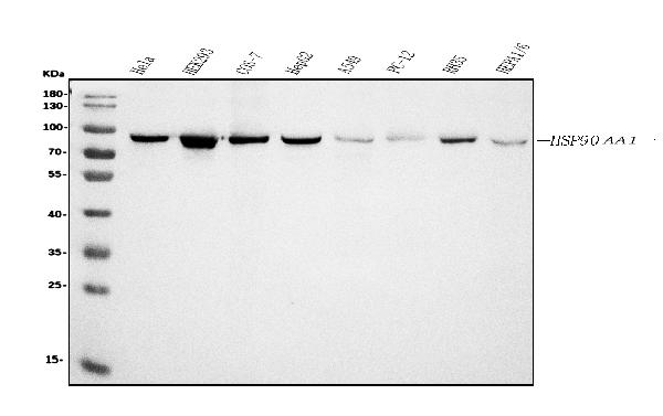
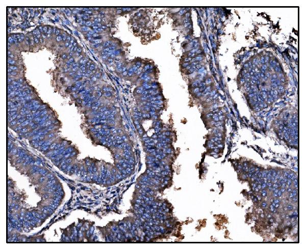
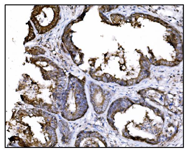
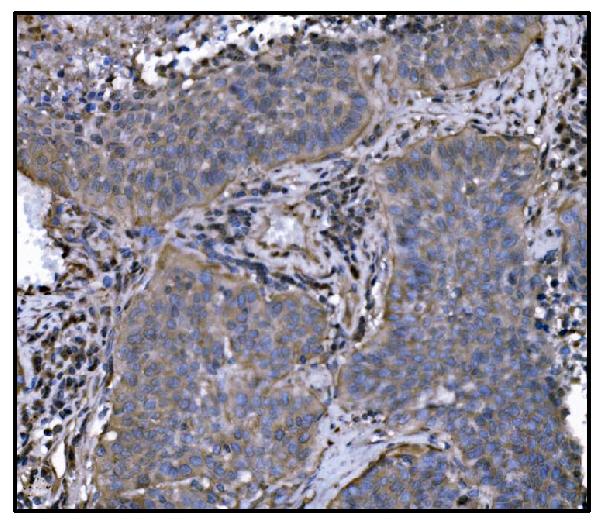
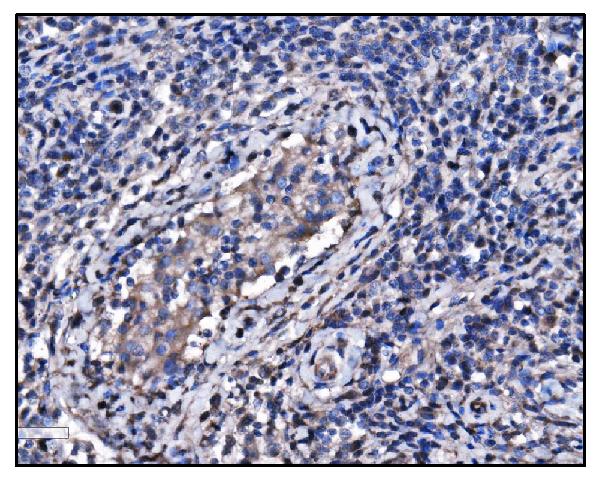
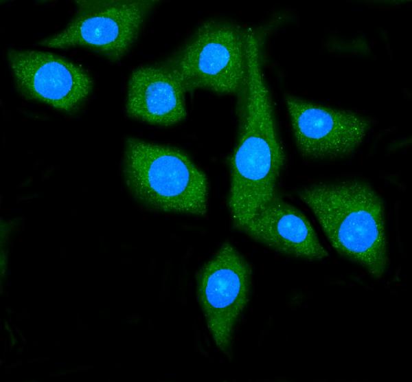
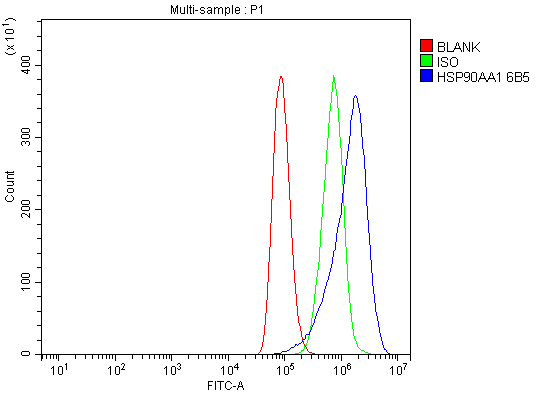
- 产品详情
- 实验流程
- 背景知识
Application 
| WB, IHC, IF, ICC, FC |
|---|---|
| Primary Accession | P07900 |
| Host | Mouse |
| Isotype | Mouse IgG2b |
| Reactivity | Rat, Human, Mouse, Monkey |
| Clonality | Monoclonal |
| Format | Lyophilized |
| Description | Anti-Hsp90 alpha Antibody Picoband™ (monoclonal, 6B5) . Tested in Flow Cytometry, IF, IHC, ICC, WB applications. This antibody reacts with Human, Monkey, Mouse, Rat. |
| Reconstitution | Add 0.2ml of distilled water will yield a concentration of 500ug/ml. |
| Gene ID | 3320 |
|---|---|
| Other Names | Heat shock protein HSP 90-alpha, 3.6.4.10, Heat shock 86 kDa, HSP 86, HSP86, Heat shock protein family C member 1, Lipopolysaccharide-associated protein 2, LAP-2, LPS-associated protein 2, Renal carcinoma antigen NY-REN-38, HSP90AA1 (HGNC:5253), HSP90A, HSPC1, HSPCA |
| Calculated MW | 84660 Da |
| Application Details | Western blot, 0.25-0.5 µg/ml, Human, Mouse, Rat, Monkey Immunohistochemistry (Paraffin-embedded Section), 2-5 µg/ml, Human Immunocytochemistry/Immunofluorescence, 5 µg/ml, Human Flow Cytometry, 1-3 µg/1x10^6 cells, Human |
| Source | Eukaryota |
| Contents | Each vial contains 4mg Trehalose, 0.9mg NaCl and 0.2mg Na2HPO4. |
| Clone Names | Clone: 6B5 |
| Immunogen | A synthetic peptide corresponding to a sequence at the C-terminus of human Hsp90 alpha, identical to the related mouse and rat sequences. |
| Purification | Immunogen affinity purified. |
| Storage | Store at -20˚C for one year from date of receipt. After reconstitution, at 4˚C for one month. It can also be aliquotted and stored frozen at -20˚C for six months. Avoid repeated freeze-thaw cycles. |
For Research Use Only. Not For Use In Diagnostic Procedures.
| Name | HSP90AA1 (HGNC:5253) |
|---|---|
| Synonyms | HSP90A, HSPC1, HSPCA |
| Function | Molecular chaperone that promotes the maturation, structural maintenance and proper regulation of specific target proteins involved for instance in cell cycle control and signal transduction. Undergoes a functional cycle that is linked to its ATPase activity which is essential for its chaperone activity. This cycle probably induces conformational changes in the client proteins, thereby causing their activation. Interacts dynamically with various co-chaperones that modulate its substrate recognition, ATPase cycle and chaperone function (PubMed:11274138, PubMed:12526792, PubMed:15577939, PubMed:15937123, PubMed:27353360, PubMed:29127155). Engages with a range of client protein classes via its interaction with various co-chaperone proteins or complexes, that act as adapters, simultaneously able to interact with the specific client and the central chaperone itself (PubMed:29127155). Recruitment of ATP and co-chaperone followed by client protein forms a functional chaperone. After the completion of the chaperoning process, properly folded client protein and co- chaperone leave HSP90 in an ADP-bound partially open conformation and finally, ADP is released from HSP90 which acquires an open conformation for the next cycle (PubMed:26991466, PubMed:27295069). Plays a critical role in mitochondrial import, delivers preproteins to the mitochondrial import receptor TOMM70 (PubMed:12526792). Apart from its chaperone activity, it also plays a role in the regulation of the transcription machinery. HSP90 and its co-chaperones modulate transcription at least at three different levels (PubMed:25973397). In the first place, they alter the steady-state levels of certain transcription factors in response to various physiological cues (PubMed:25973397). Second, they modulate the activity of certain epigenetic modifiers, such as histone deacetylases or DNA methyl transferases, and thereby respond to the change in the environment (PubMed:25973397). Third, they participate in the eviction of histones from the promoter region of certain genes and thereby turn on gene expression (PubMed:25973397). Binds bacterial lipopolysaccharide (LPS) and mediates LPS-induced inflammatory response, including TNF secretion by monocytes (PubMed:11276205). Antagonizes STUB1-mediated inhibition of TGF-beta signaling via inhibition of STUB1-mediated SMAD3 ubiquitination and degradation (PubMed:24613385). Mediates the association of TOMM70 with IRF3 or TBK1 in mitochondrial outer membrane which promotes host antiviral response (PubMed:20628368, PubMed:25609812). |
| Cellular Location | Nucleus {ECO:0000250|UniProtKB:P07901}. Cytoplasm {ECO:0000250|UniProtKB:P07901}. Melanosome. Cell membrane. Mitochondrion. Note=Identified by mass spectrometry in melanosome fractions from stage I to stage IV |
Provided below are standard protocols that you may find useful for product applications.
BACKGROUND
Heat shock protein HSP 90-alpha is a protein that in humans is encoded by the HSP90AA1 gene. The gene, HSP90AA1, encodes the human stress-inducible 90-kDa heat shock protein alpha (Hsp90A). Complemented by the constitutively expressed paralog Hsp90B which shares over 85% amino acid sequence identity, Hsp90A expression is initiated when a cell experiences proteotoxic stress. Once expressed Hsp90A dimers operate as molecular chaperones that bind and fold other proteins into their functional 3-dimensional structures. This molecular chaperoning ability of Hsp90A is driven by a cycle of structural rearrangements fueled by ATP hydrolysis. Current research on Hsp90A focuses in its role as a drug target due to its interaction with a large number of tumor promoting proteins and its role in cellular stress adaptation.

终于等到您。ABCEPTA(百远生物)抗体产品。
点击下方“我要评价 ”按钮提交您的反馈信息,您的反馈和评价是我们最宝贵的财富之一,
我们将在1-3个工作日内处理您的反馈信息。
如有疑问,联系:0512-88856768 tech-china@abcepta.com.


