


 癌症的基本特征包括细胞增殖、血管生成、迁移、凋亡逃避机制和细胞永生等。找到癌症发生过程中这些通路的关键标记物和对应的抗体用于检测至关重要。
 为您推荐一个泛素化位点预测神器——泛素化分析工具,可以为您的蛋白的泛素化位点作出预测和评分。
 细胞自噬受体图形绘图工具为你的蛋白的细胞受体结合位点作出预测和评分,识别结合到自噬通路中的蛋白是非常重要的,便于让我们理解自噬在正常生理、病理过程中的作用,如发育、细胞分化、神经退化性疾病、压力条件下、感染和癌症。
Anti-ITCH/AIP4 Antibody Picoband™ (monoclonal, 5E12)
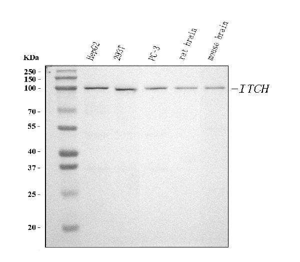
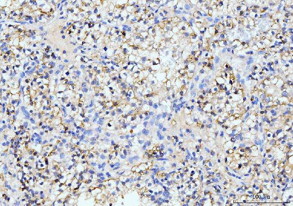
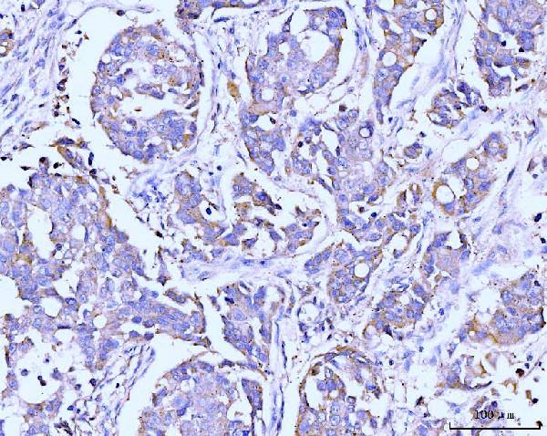
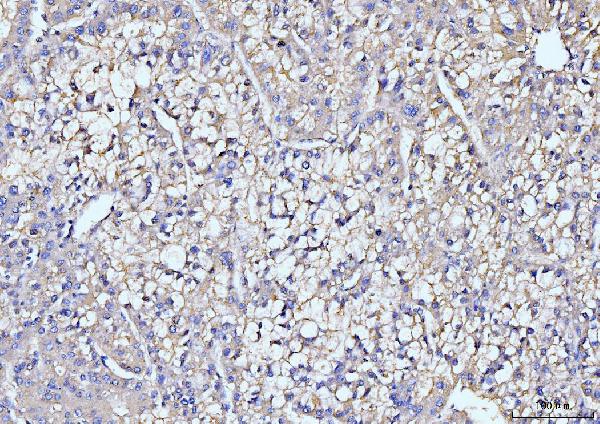
- 产品详情
- 实验流程
- 背景知识
Application 
| WB, IHC, FC |
|---|---|
| Primary Accession | Q96J02 |
| Host | Mouse |
| Isotype | Mouse IgG2b |
| Reactivity | Rat, Human, Mouse |
| Clonality | Monoclonal |
| Format | Lyophilized |
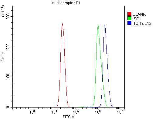
| Description | Anti-ITCH/AIP4 Antibody Picoband™ (monoclonal, 5E12) . Tested in FCM, IHC, WB applications. This antibody reacts with Human, Mouse, Rat. |
| Reconstitution | Adding 0.2 ml of distilled water will yield a concentration of 500 µg/ml. |
| Gene ID | 83737 |
|---|---|
| Other Names | E3 ubiquitin-protein ligase Itchy homolog, Itch, 2.3.2.26, Atrophin-1-interacting protein 4, AIP4, HECT-type E3 ubiquitin transferase Itchy homolog, NFE2-associated polypeptide 1, NAPP1, ITCH |
| Calculated MW | 102803 Da |
| Application Details | Western blot, 0.25-0.5 µg/ml, Human, Mouse, Rat Immunohistochemistry(Paraffin-embedded Section), 2-5 µg/ml, Human Flow Cytometry, 1-3 µg/1x^6 cells, Human |
| Source | Eukaryota |
| Contents | Each vial contains 4 mg Trehalose, 0.9 mg NaCl and 0.2 mg Na2HPO4. |
| Clone Names | Clone: 5E12 |
| Immunogen | E.coli-derived human ITCH/AIP4 recombinant protein (Position: K17-E358). |
| Purification | Immunogen affinity purified. |
| Storage | At -20°C for one year from date of receipt. After reconstitution, at 4°C for one month. It can also be aliquotted and stored frozen at -20°C for six months. Avoid repeated freezing and thawing. |
For Research Use Only. Not For Use In Diagnostic Procedures.
| Name | ITCH |
|---|---|
| Function | Acts as an Acts as an E3 ubiquitin-protein ligase which accepts ubiquitin from an E2 ubiquitin-conjugating enzyme in the form of a thioester and then directly transfers the ubiquitin to targeted substrates (PubMed:11046148, PubMed:14602072, PubMed:15051726, PubMed:16387660, PubMed:17028573, PubMed:18718448, PubMed:18718449, PubMed:19116316, PubMed:19592251, PubMed:19881509, PubMed:20068034, PubMed:20392206, PubMed:20491914, PubMed:23146885, PubMed:24790097, PubMed:25631046). Catalyzes 'Lys-29'-, 'Lys-48'- and 'Lys-63'-linked ubiquitin conjugation (PubMed:17028573, PubMed:18718448, PubMed:19131965, PubMed:19881509). Involved in the control of inflammatory signaling pathways (PubMed:19131965). Essential component of a ubiquitin-editing protein complex, comprising also TNFAIP3, TAX1BP1 and RNF11, that ensures the transient nature of inflammatory signaling pathways (PubMed:19131965). Promotes the association of the complex after TNF stimulation (PubMed:19131965). Once the complex is formed, TNFAIP3 deubiquitinates 'Lys-63' polyubiquitin chains on RIPK1 and catalyzes the formation of 'Lys-48'-polyubiquitin chains (PubMed:19131965). This leads to RIPK1 proteasomal degradation and consequently termination of the TNF- or LPS-mediated activation of NFKB1 (PubMed:19131965). Ubiquitinates RIPK2 by 'Lys-63'-linked conjugation and influences NOD2-dependent signal transduction pathways (PubMed:19592251). Regulates the transcriptional activity of several transcription factors, and probably plays an important role in the regulation of immune response (PubMed:18718448, PubMed:20491914). Ubiquitinates NFE2 by 'Lys-63' linkages and is implicated in the control of the development of hematopoietic lineages (PubMed:18718448). Mediates JUN ubiquitination and degradation (By similarity). Mediates JUNB ubiquitination and degradation (PubMed:16387660). Critical regulator of type 2 helper T (Th2) cell cytokine production by inducing JUNB ubiquitination and degradation (By similarity). Involved in the negative regulation of MAVS-dependent cellular antiviral responses (PubMed:19881509). Ubiquitinates MAVS through 'Lys-48'-linked conjugation resulting in MAVS proteasomal degradation (PubMed:19881509). Following ligand stimulation, regulates sorting of Wnt receptor FZD4 to the degradative endocytic pathway probably by modulating PI42KA activity (PubMed:23146885). Ubiquitinates PI4K2A and negatively regulates its catalytic activity (PubMed:23146885). Ubiquitinates chemokine receptor CXCR4 and regulates sorting of CXCR4 to the degradative endocytic pathway following ligand stimulation by ubiquitinating endosomal sorting complex required for transport ESCRT-0 components HGS and STAM (PubMed:14602072, PubMed:23146885, PubMed:34927784). Targets DTX1 for lysosomal degradation and controls NOTCH1 degradation, in the absence of ligand, through 'Lys-29'-linked polyubiquitination (PubMed:17028573, PubMed:18628966, PubMed:23886940). Ubiquitinates SNX9 (PubMed:20491914). Ubiquitinates MAP3K7 through 'Lys-48'-linked conjugation (By similarity). Together with UBR5, involved in the regulation of apoptosis and reactive oxygen species levels through the ubiquitination and proteasomal degradation of TXNIP: catalyzes 'Lys-48'-/'Lys-63'-branched ubiquitination of TXNIP (PubMed:20068034, PubMed:29378950). ITCH synthesizes 'Lys-63'-linked chains, while UBR5 is branching multiple 'Lys-48'-linked chains of substrate initially modified (PubMed:29378950). Mediates the antiapoptotic activity of epidermal growth factor through the ubiquitination and proteasomal degradation of p15 BID (PubMed:20392206). Ubiquitinates BRAT1 and this ubiquitination is enhanced in the presence of NDFIP1 (PubMed:25631046). Inhibits the replication of influenza A virus (IAV) via ubiquitination of IAV matrix protein 1 (M1) through 'Lys-48'-linked conjugation resulting in M1 proteasomal degradation (PubMed:30328013). Ubiquitinates NEDD9/HEF1, resulting in proteasomal degradation of NEDD9/HEF1 (PubMed:15051726). |
| Cellular Location | Cell membrane; Peripheral membrane protein; Cytoplasmic side. Cytoplasm. Nucleus Early endosome membrane; Peripheral membrane protein; Cytoplasmic side. Endosome membrane; Peripheral membrane protein; Cytoplasmic side. Note=May be recruited to exosomes by NDFIP1 (PubMed:18819914). Localizes to plasma membrane upon CXCL12 stimulation where it co-localizes with CXCL4 (PubMed:14602072) Localization to early endosomes is increased upon CXCL12 stimulation where it co-localizes with DTX3L and CXCL4 (PubMed:24790097) |
| Tissue Location | Widely expressed. |
Provided below are standard protocols that you may find useful for product applications.
BACKGROUND
ITCH is an ubiquitin-conjugating enzyme. This gene encodes a member of the Nedd4 family of HECT domain E3 ubiquitin ligases. HECT domain E3 ubiquitin ligases transfer ubiquitin from E2 ubiquitin-conjugating enzymes to protein substrates, thus targeting specific proteins for lysosomal degradation. The encoded protein plays a role in multiple cellular processes including erythroid and lymphoid cell differentiation and the regulation of immune responses. Mutations in this gene are a cause of syndromic multisystem autoimmune disease. Alternatively spliced transcript variants encoding multiple isoforms have been observed for this gene.

终于等到您。ABCEPTA(百远生物)抗体产品。
点击下方“我要评价 ”按钮提交您的反馈信息,您的反馈和评价是我们最宝贵的财富之一,
我们将在1-3个工作日内处理您的反馈信息。
如有疑问,联系:0512-88856768 tech-china@abcepta.com.


