


 癌症的基本特征包括细胞增殖、血管生成、迁移、凋亡逃避机制和细胞永生等。找到癌症发生过程中这些通路的关键标记物和对应的抗体用于检测至关重要。
 为您推荐一个泛素化位点预测神器——泛素化分析工具,可以为您的蛋白的泛素化位点作出预测和评分。
 细胞自噬受体图形绘图工具为你的蛋白的细胞受体结合位点作出预测和评分,识别结合到自噬通路中的蛋白是非常重要的,便于让我们理解自噬在正常生理、病理过程中的作用,如发育、细胞分化、神经退化性疾病、压力条件下、感染和癌症。
Anti-RACK1 Rabbit Monoclonal Antibody
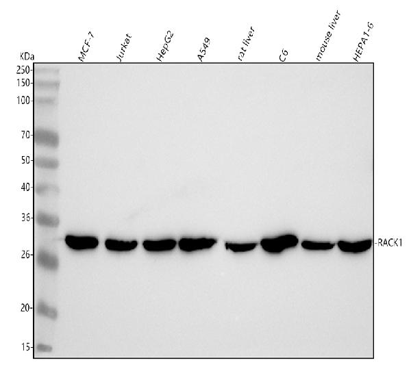
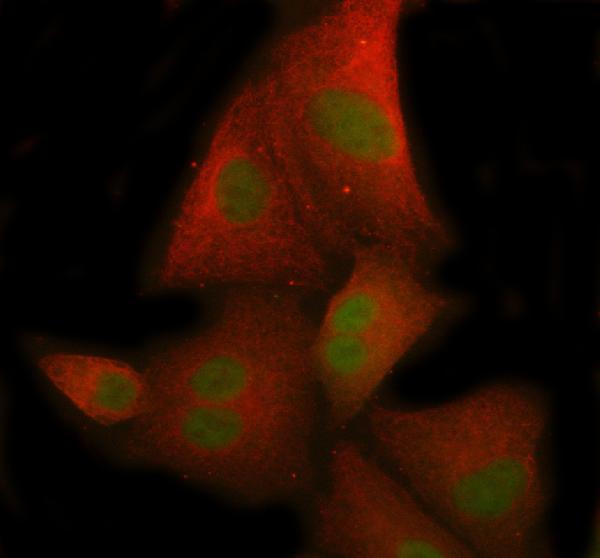
- 产品详情
- 实验流程
Application 
| WB, IHC, IF, ICC, FC |
|---|---|
| Primary Accession | P63244 |
| Host | Rabbit |
| Isotype | IgG |
| Reactivity | Rat, Human, Mouse |
| Clonality | Monoclonal |
| Format | Liquid |
| Description | Anti-RACK1 Rabbit Monoclonal Antibody . Tested in WB, IHC, ICC/IF, Flow Cytometry applications. This antibody reacts with Human, Mouse, Rat. |
| Gene ID | 10399 |
|---|---|
| Other Names | Small ribosomal subunit protein RACK1, Cell proliferation-inducing gene 21 protein, Guanine nucleotide-binding protein subunit beta-2-like 1, Guanine nucleotide-binding protein subunit beta-like protein 12.3, Human lung cancer oncogene 7 protein, HLC-7, Receptor for activated C kinase, Receptor of activated protein C kinase 1, Small ribosomal subunit protein RACK1, N-terminally processed, Guanine nucleotide-binding protein subunit beta-2-like 1, N-terminally processed, Receptor of activated protein C kinase 1, N-terminally processed, RACK1 (HGNC:4399), GNB2L1 |
| Calculated MW | 35077 Da |
| Application Details | WB 1:500-1:2000 IHC 1:50-1:200 ICC/IF 1:50-1:200 FC 1:50 |
| Source | Eukaryota |
| Contents | Rabbit IgG in phosphate buffered saline, pH 7.4, 150mM NaCl, 0.02% sodium azide and 50% glycerol, 0.4-0.5mg/ml BSA. |
| Clone Names | Clone: 20R31 |
| Immunogen | A synthesized peptide derived from human RACK1 |
| Purification | Affinity-chromatography |
| Storage | Store at -20°C for one year. For short term storage and frequent use, store at 4°C for up to one month. Avoid repeated freeze-thaw cycles. |
For Research Use Only. Not For Use In Diagnostic Procedures.
| Name | RACK1 (HGNC:4399) |
|---|---|
| Synonyms | GNB2L1 |
| Function | Scaffolding protein involved in the recruitment, assembly and/or regulation of a variety of signaling molecules. Interacts with a wide variety of proteins and plays a role in many cellular processes. Component of the 40S ribosomal subunit involved in translational repression (PubMed:23636399). Involved in the initiation of the ribosome quality control (RQC), a pathway that takes place when a ribosome has stalled during translation, by promoting ubiquitination of a subset of 40S ribosomal subunits (PubMed:28132843). Binds to and stabilizes activated protein kinase C (PKC), increasing PKC-mediated phosphorylation. May recruit activated PKC to the ribosome, leading to phosphorylation of EIF6. Inhibits the activity of SRC kinases including SRC, LCK and YES1. Inhibits cell growth by prolonging the G0/G1 phase of the cell cycle. Enhances phosphorylation of BMAL1 by PRKCA and inhibits transcriptional activity of the BMAL1-CLOCK heterodimer. Facilitates ligand-independent nuclear translocation of AR following PKC activation, represses AR transactivation activity and is required for phosphorylation of AR by SRC. Modulates IGF1R-dependent integrin signaling and promotes cell spreading and contact with the extracellular matrix. Involved in PKC-dependent translocation of ADAM12 to the cell membrane. Promotes the ubiquitination and proteasome- mediated degradation of proteins such as CLEC1B and HIF1A. Required for VANGL2 membrane localization, inhibits Wnt signaling, and regulates cellular polarization and oriented cell division during gastrulation. Required for PTK2/FAK1 phosphorylation and dephosphorylation. Regulates internalization of the muscarinic receptor CHRM2. Promotes apoptosis by increasing oligomerization of BAX and disrupting the interaction of BAX with the anti-apoptotic factor BCL2L. Inhibits TRPM6 channel activity. Regulates cell surface expression of some GPCRs such as TBXA2R. Plays a role in regulation of FLT1-mediated cell migration. Involved in the transport of ABCB4 from the Golgi to the apical bile canalicular membrane (PubMed:19674157). Promotes migration of breast carcinoma cells by binding to and activating RHOA (PubMed:20499158). Acts as an adapter for the dephosphorylation and inactivation of AKT1 by promoting recruitment of PP2A phosphatase to AKT1 (By similarity). |
| Cellular Location | Cell membrane; Peripheral membrane protein. Cytoplasm. Cytoplasm, perinuclear region. Nucleus. Perikaryon {ECO:0000250|UniProtKB:P68040}. Cell projection, dendrite {ECO:0000250|UniProtKB:P68040}. Cell projection, phagocytic cup. Note=Recruited to the plasma membrane through interaction with KRT1 which binds to membrane-bound ITGB1 (PubMed:17956333). Also associated with the membrane in oncogene- transformed cells (PubMed:11884618). PKC activation induces translocation from the perinuclear region to the cell periphery (PubMed:11279199). In the brain, detected mainly in cell bodies and dendrites with little expression in axonal fibers or nuclei (By similarity). Localized to phagocytic cups following infection by Y.pestis (PubMed:21347310). {ECO:0000250|UniProtKB:P68040, ECO:0000269|PubMed:11279199, ECO:0000269|PubMed:11884618, ECO:0000269|PubMed:17956333, ECO:0000269|PubMed:21347310} |
| Tissue Location | In the liver, expressed at higher levels in activated hepatic stellate cells than in hepatocytes or Kupffer cells Up-regulated in hepatocellular carcinomas and in the adjacent non-tumor liver tissue. |
Research Areas
Application Protocols
Provided below are standard protocols that you may find useful for product applications.

终于等到您。ABCEPTA(百远生物)抗体产品。
点击下方“我要评价 ”按钮提交您的反馈信息,您的反馈和评价是我们最宝贵的财富之一,
我们将在1-3个工作日内处理您的反馈信息。
如有疑问,联系:0512-88856768 tech-china@abcepta.com.


