注册 | 登录
点击这里给我发消息
所有产品
  • 所有产品
  • 一抗
  • 裂解液
>   首页   >   产品   >   一抗   >   信号转导   >   Anti-QK1 Rabbit Monoclonal Antibody   
Anti-QK1 Rabbit Monoclonal Antibody
     
  • 1 - Anti-QK1 Rabbit Monoclonal Antibody ABO16077
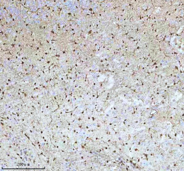
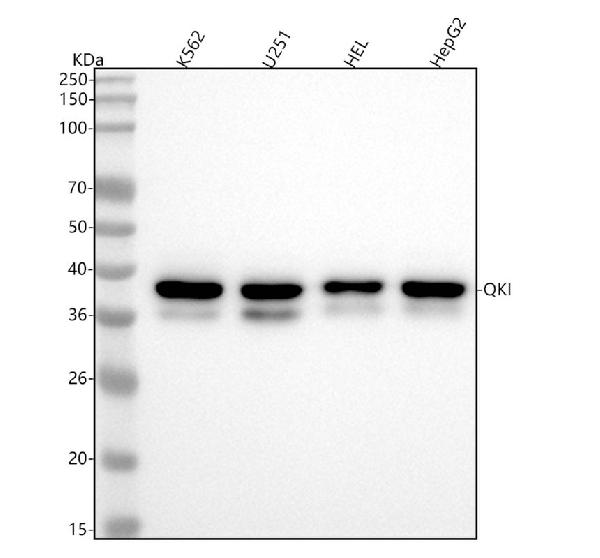
    Figure 1. Western blot analysis of QK1 using anti-QK1 antibody (M01874).
    Electrophoresis was performed on a 5-20% SDS-PAGE gel at 70V (Stacking gel) / 90V (Resolving gel) for 2-3 hours. The sample well of each lane was loaded with 30 ug of sample under reducing conditions.
    Lane 1: human K562 whole cell lysates,
    Lane 2: human U251 whole cell lysates,
    Lane 3: human HEL whole cell lysates,
    Lane 4: human HepG2 whole cell lysates.
    After electrophoresis, proteins were transferred to a nitrocellulose membrane at 150 mA for 50-90 minutes. Blocked the membrane with 5% non-fat milk/TBS for 1.5 hour at RT. The membrane was incubated with rabbit anti-QK1 antigen affinity purified monoclonal antibody (Catalog # M01874) at 1:500 overnight at 4°C, then washed with TBS-0.1%Tween 3 times with 5 minutes each and probed with a goat anti-rabbit IgG-HRP secondary antibody at a dilution of 1:5000 for 1.5 hour at RT. The signal is developed using an Enhanced Chemiluminescent detection (ECL) kit (Catalog # EK1002) with Tanon 5200 system. A specific band was detected for QK1 at approximately 38 kDa. The expected band size for QK1 is at 38 kDa.
  • 2 - Anti-QK1 Rabbit Monoclonal Antibody ABO16077
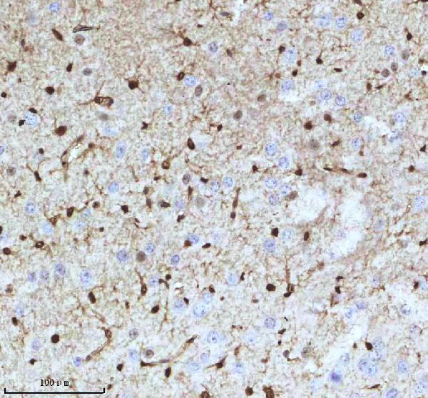
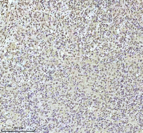
    Figure 2. IHC analysis of QK1 using anti-QK1 antibody (M01874).
    QK1 was detected in a paraffin-embedded section of human glioma tissue. Heat mediated antigen retrieval was performed in EDTA buffer (pH 8.0, epitope retrieval solution). The tissue section was blocked with 10% goat serum. The tissue section was then incubated with 1:50 rabbit anti-QK1 Antibody (M01874) overnight at 4°C. Peroxidase Conjugated Goat Anti-rabbit IgG was used as secondary antibody and incubated for 30 minutes at 37°C. The tissue section was developed using HRP Conjugated Rabbit IgG Super Vision Assay Kit (Catalog # SV0002) with DAB as the chromogen.
  • 2 - Anti-QK1 Rabbit Monoclonal Antibody ABO16077
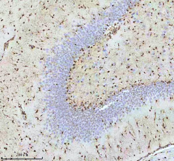
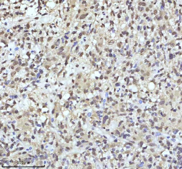
    Figure 3. IHC analysis of QK1 using anti-QK1 antibody (M01874).
    QK1 was detected in a paraffin-embedded section of human glioma tissue. Heat mediated antigen retrieval was performed in EDTA buffer (pH 8.0, epitope retrieval solution). The tissue section was blocked with 10% goat serum. The tissue section was then incubated with 1:50 rabbit anti-QK1 Antibody (M01874) overnight at 4°C. Peroxidase Conjugated Goat Anti-rabbit IgG was used as secondary antibody and incubated for 30 minutes at 37°C. The tissue section was developed using HRP Conjugated Rabbit IgG Super Vision Assay Kit (Catalog # SV0002) with DAB as the chromogen.
  • 2 - Anti-QK1 Rabbit Monoclonal Antibody ABO16077
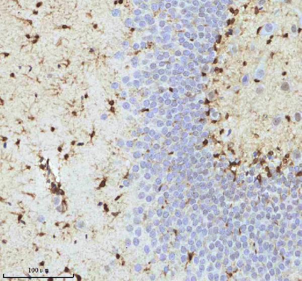
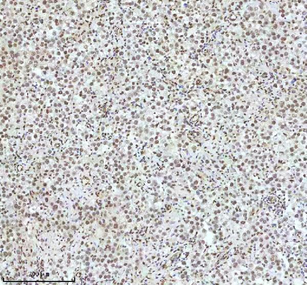
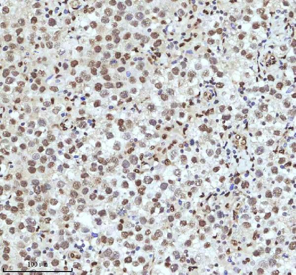
    Figure 4. IHC analysis of QK1 using anti-QK1 antibody (M01874).
    QK1 was detected in a paraffin-embedded section of human testis cancer tissue. Heat mediated antigen retrieval was performed in EDTA buffer (pH 8.0, epitope retrieval solution). The tissue section was blocked with 10% goat serum. The tissue section was then incubated with 1:50 rabbit anti-QK1 Antibody (M01874) overnight at 4°C. Peroxidase Conjugated Goat Anti-rabbit IgG was used as secondary antibody and incubated for 30 minutes at 37°C. The tissue section was developed using HRP Conjugated Rabbit IgG Super Vision Assay Kit (Catalog # SV0002) with DAB as the chromogen.
  • 2 - Anti-QK1 Rabbit Monoclonal Antibody ABO16077
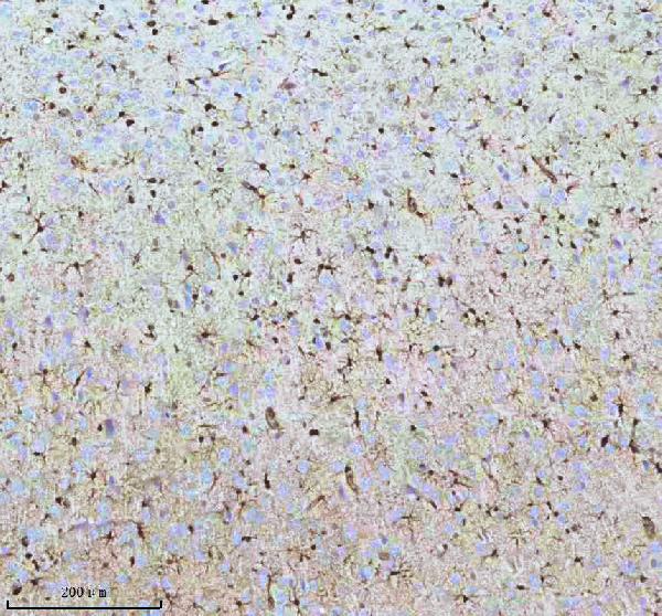
    Figure 5. IHC analysis of QK1 using anti-QK1 antibody (M01874).
    QK1 was detected in a paraffin-embedded section of human testis cancer tissue. Heat mediated antigen retrieval was performed in EDTA buffer (pH 8.0, epitope retrieval solution). The tissue section was blocked with 10% goat serum. The tissue section was then incubated with 1:50 rabbit anti-QK1 Antibody (M01874) overnight at 4°C. Peroxidase Conjugated Goat Anti-rabbit IgG was used as secondary antibody and incubated for 30 minutes at 37°C. The tissue section was developed using HRP Conjugated Rabbit IgG Super Vision Assay Kit (Catalog # SV0002) with DAB as the chromogen.
  • 2 - Anti-QK1 Rabbit Monoclonal Antibody ABO16077
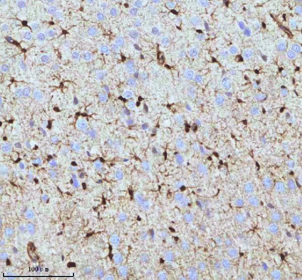
    Figure 6. IHC analysis of QK1 using anti-QK1 antibody (M01874).
    QK1 was detected in a paraffin-embedded section of mouse brain tissue. Heat mediated antigen retrieval was performed in EDTA buffer (pH 8.0, epitope retrieval solution). The tissue section was blocked with 10% goat serum. The tissue section was then incubated with 1:50 rabbit anti-QK1 Antibody (M01874) overnight at 4°C. Peroxidase Conjugated Goat Anti-rabbit IgG was used as secondary antibody and incubated for 30 minutes at 37°C. The tissue section was developed using HRP Conjugated Rabbit IgG Super Vision Assay Kit (Catalog # SV0002) with DAB as the chromogen.
  • 2 - Anti-QK1 Rabbit Monoclonal Antibody ABO16077
    Figure 7. IHC analysis of QK1 using anti-QK1 antibody (M01874).
    QK1 was detected in a paraffin-embedded section of mouse brain tissue. Heat mediated antigen retrieval was performed in EDTA buffer (pH 8.0, epitope retrieval solution). The tissue section was blocked with 10% goat serum. The tissue section was then incubated with 1:50 rabbit anti-QK1 Antibody (M01874) overnight at 4°C. Peroxidase Conjugated Goat Anti-rabbit IgG was used as secondary antibody and incubated for 30 minutes at 37°C. The tissue section was developed using HRP Conjugated Rabbit IgG Super Vision Assay Kit (Catalog # SV0002) with DAB as the chromogen.
  • 2 - Anti-QK1 Rabbit Monoclonal Antibody ABO16077
    Figure 8. IHC analysis of QK1 using anti-QK1 antibody (M01874).
    QK1 was detected in a paraffin-embedded section of rat brain tissue. Heat mediated antigen retrieval was performed in EDTA buffer (pH 8.0, epitope retrieval solution). The tissue section was blocked with 10% goat serum. The tissue section was then incubated with 1:50 rabbit anti-QK1 Antibody (M01874) overnight at 4°C. Peroxidase Conjugated Goat Anti-rabbit IgG was used as secondary antibody and incubated for 30 minutes at 37°C. The tissue section was developed using HRP Conjugated Rabbit IgG Super Vision Assay Kit (Catalog # SV0002) with DAB as the chromogen.
  • 2 - Anti-QK1 Rabbit Monoclonal Antibody ABO16077
    Figure 9. IHC analysis of QK1 using anti-QK1 antibody (M01874).
    QK1 was detected in a paraffin-embedded section of rat brain tissue. Heat mediated antigen retrieval was performed in EDTA buffer (pH 8.0, epitope retrieval solution). The tissue section was blocked with 10% goat serum. The tissue section was then incubated with 1:50 rabbit anti-QK1 Antibody (M01874) overnight at 4°C. Peroxidase Conjugated Goat Anti-rabbit IgG was used as secondary antibody and incubated for 30 minutes at 37°C. The tissue section was developed using HRP Conjugated Rabbit IgG Super Vision Assay Kit (Catalog # SV0002) with DAB as the chromogen.
  • 2 - Anti-QK1 Rabbit Monoclonal Antibody ABO16077
    Figure 10. IHC analysis of QK1 using anti-QK1 antibody (M01874).
    QK1 was detected in a paraffin-embedded section of rat brain tissue. Heat mediated antigen retrieval was performed in EDTA buffer (pH 8.0, epitope retrieval solution). The tissue section was blocked with 10% goat serum. The tissue section was then incubated with 1:50 rabbit anti-QK1 Antibody (M01874) overnight at 4°C. Peroxidase Conjugated Goat Anti-rabbit IgG was used as secondary antibody and incubated for 30 minutes at 37°C. The tissue section was developed using HRP Conjugated Rabbit IgG Super Vision Assay Kit (Catalog # SV0002) with DAB as the chromogen.
  • 2 - Anti-QK1 Rabbit Monoclonal Antibody ABO16077
    Figure 11. IHC analysis of QK1 using anti-QK1 antibody (M01874).
    QK1 was detected in a paraffin-embedded section of rat brain tissue. Heat mediated antigen retrieval was performed in EDTA buffer (pH 8.0, epitope retrieval solution). The tissue section was blocked with 10% goat serum. The tissue section was then incubated with 1:50 rabbit anti-QK1 Antibody (M01874) overnight at 4°C. Peroxidase Conjugated Goat Anti-rabbit IgG was used as secondary antibody and incubated for 30 minutes at 37°C. The tissue section was developed using HRP Conjugated Rabbit IgG Super Vision Assay Kit (Catalog # SV0002) with DAB as the chromogen.
  • 产品详情
  • 实验流程
Product Information
Application
  • Applications Legend:
  • E=ELISA
  • WB=Western Blotting
  • IHC=Immunohistochemistry
  • IHC-P=Immunohistochemistry (Paraffin)
  • IP=Immunoprecipitation
  • IF=Immunofluorescence
  • IC=Immunochemistry
  • ICC=Immunocytochemistry
  • FC=Flow Cytometry
  • DB=Dot Blot
WB, IHC, IF, ICC, IP
Primary Accession Q96PU8
Host Rabbit
Isotype IgG
Reactivity Rat, Human, Mouse
Clonality Monoclonal
Format Liquid
Description Anti-QK1 Rabbit Monoclonal Antibody . Tested in WB, IHC, ICC/IF, IP applications. This antibody reacts with Human, Mouse.
Additional Information
Gene ID 9444
Other Names KH domain-containing RNA-binding protein QKI, Protein quaking, Hqk, HqkI, QKI {ECO:0000303|PubMed:16342280, ECO:0000312|HGNC:HGNC:21100}
Calculated MW 37671 Da
Application Details WB 1:500-1:2000
IHC 1:50-1:200
ICC/IF 1:50-1:200
IP 1:50
Source Eukaryota
Contents Rabbit IgG in phosphate buffered saline, pH 7.4, 150mM NaCl, 0.02% sodium azide and 50% glycerol, 0.4-0.5mg/ml BSA.
Clone Names Clone: 26Q56
Immunogen A synthesized peptide derived from QK1
Purification Affinity-chromatography
Storage Store at -20°C for one year. For short term storage and frequent use, store at 4°C for up to one month. Avoid repeated freeze-thaw cycles.

For Research Use Only. Not For Use In Diagnostic Procedures.

Protein Information
Name QKI {ECO:0000303|PubMed:16342280, ECO:0000312|HGNC:HGNC:21100}
Function RNA reader protein, which recognizes and binds specific RNAs, thereby regulating RNA metabolic processes, such as pre-mRNA splicing, circular RNA (circRNA) formation, mRNA export, mRNA stability and/or translation (PubMed:22398723, PubMed:23630077, PubMed:25768908, PubMed:27029405, PubMed:31331967, PubMed:37379838). Involved in various cellular processes, such as mRNA storage into stress granules, apoptosis, lipid deposition, interferon response, glial cell fate and development (PubMed:25768908, PubMed:31829086, PubMed:34428287, PubMed:37379838). Binds to the 5'-NACUAAY-N(1,20)-UAAY-3' RNA core sequence (PubMed:23630077). Acts as a mRNA modification reader that specifically recognizes and binds mRNA transcripts modified by internal N(7)-methylguanine (m7G) (PubMed:37379838). Promotes the formation of circular RNAs (circRNAs) during the epithelial to mesenchymal transition and in cardiomyocytes: acts by binding to sites flanking circRNA-forming exons (PubMed:25768908). CircRNAs are produced by back- splicing circularization of pre-mRNAs (PubMed:25768908). Plays a central role in myelinization via 3 distinct mechanisms (PubMed:16641098). First, acts by protecting and promoting stability of target mRNAs such as MBP, SIRT2 and CDKN1B, which promotes oligodendrocyte differentiation (By similarity). Second, participates in mRNA transport by regulating the nuclear export of MBP mRNA (By similarity). Finally, indirectly regulates mRNA splicing of MAG pre- mRNA during oligodendrocyte differentiation by acting as a negative regulator of MAG exon 12 alternative splicing: acts by binding to HNRNPA1 mRNA splicing factor, preventing its translation (By similarity). Involved in microglia differentiation and remyelination by regulating microexon alternative splicing of the Rho GTPase pathway (By similarity). Involved in macrophage differentiation: promotes monocyte differentiation by regulating pre-mRNA splicing in naive peripheral blood monocytes (PubMed:27029405). Acts as an important regulator of muscle development: required for the contractile function of cardiomyocytes by regulating alternative splicing of cardiomyocyte transcripts (By similarity). Acts as a negative regulator of thermogenesis by decreasing stability, nuclear export and translation of mRNAs encoding PPARGC1A and UCP1 (By similarity). Also required for visceral endoderm function and blood vessel development (By similarity). May also play a role in smooth muscle development (PubMed:31331967). In addition to its RNA-binding activity, also acts as a nuclear transcription coactivator for SREBF2/SREBP2 (By similarity).
Cellular Location Nucleus. Cytoplasm [Isoform QKI6]: Cytoplasm, cytosol. Nucleus Note=Localizes predominantly in the cytoplasm and at lower levels in nucleus.
Tissue Location Expressed in the frontal cortex of brain. Down- regulated in the brain of schizophrenic patients
Research Areas
FeedBack

终于等到您。ABCEPTA(百远生物)抗体产品。
点击下方“我要评价 ”按钮提交您的反馈信息,您的反馈和评价是我们最宝贵的财富之一,
我们将在1-3个工作日内处理您的反馈信息。

如有疑问,联系:0512-88856768 tech-china@abcepta.com.


我要评价