注册 | 登录
点击这里给我发消息
所有产品
  • 所有产品
  • 一抗
  • 裂解液
>   首页   >   产品   >   一抗   >   信号转导   >   Anti-HNRNPM Rabbit Monoclonal Antibody   
Anti-HNRNPM Rabbit Monoclonal Antibody
     
  • 1 - Anti-HNRNPM Rabbit Monoclonal Antibody ABO16441
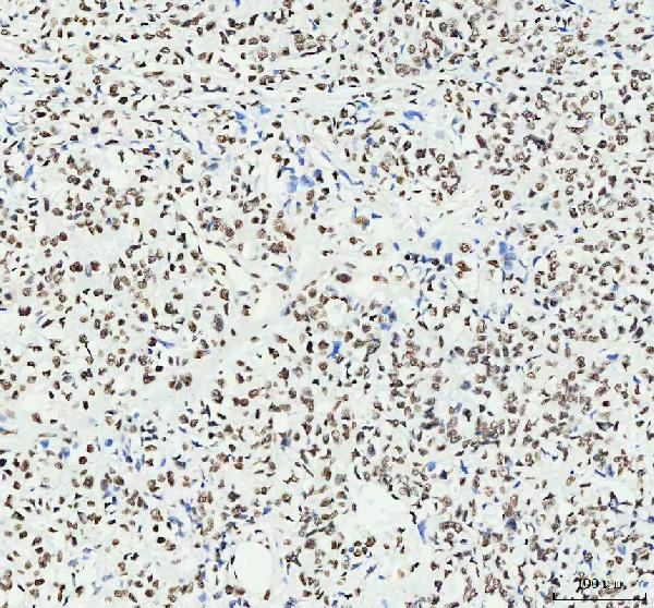
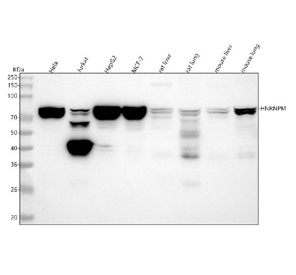
    Figure 1. Western blot analysis of HNRNPM using anti-HNRNPM antibody (M06017-1).
    Electrophoresis was performed on a 5-20% SDS-PAGE gel at 70V (Stacking gel) / 90V (Resolving gel) for 2-3 hours. The sample well of each lane was loaded with 30 ug of sample under reducing conditions.
    Lane 1: human Hela whole cell lysates,
    Lane 2: human Jurkat whole cell lysates,
    Lane 3: human HepG2 whole cell lysates,
    Lane 4: human MCF-7 whole cell lysates,
    Lane 5: rat liver tissue lysates,
    Lane 6: rat lung tissue lysates,
    Lane 7: mouse liver tissue lysates,
    Lane 8: mouse lung tissue lysates.
    After electrophoresis, proteins were transferred to a nitrocellulose membrane at 150 mA for 50-90 minutes. Blocked the membrane with 5% non-fat milk/TBS for 1.5 hour at RT. The membrane was incubated with rabbit anti-HNRNPM antigen affinity purified monoclonal antibody (Catalog # M06017-1) at 1:500 overnight at 4°C, then washed with TBS-0.1%Tween 3 times with 5 minutes each and probed with a goat anti-rabbit IgG-HRP secondary antibody at a dilution of 1:5000 for 1.5 hour at RT. The signal is developed using an Enhanced Chemiluminescent detection (ECL) kit (Catalog # EK1002) with Tanon 5200 system. A specific band was detected for HNRNPM at approximately 73 kDa. The expected band size for HNRNPM is at 76 kDa.
  • 2 - Anti-HNRNPM Rabbit Monoclonal Antibody ABO16441
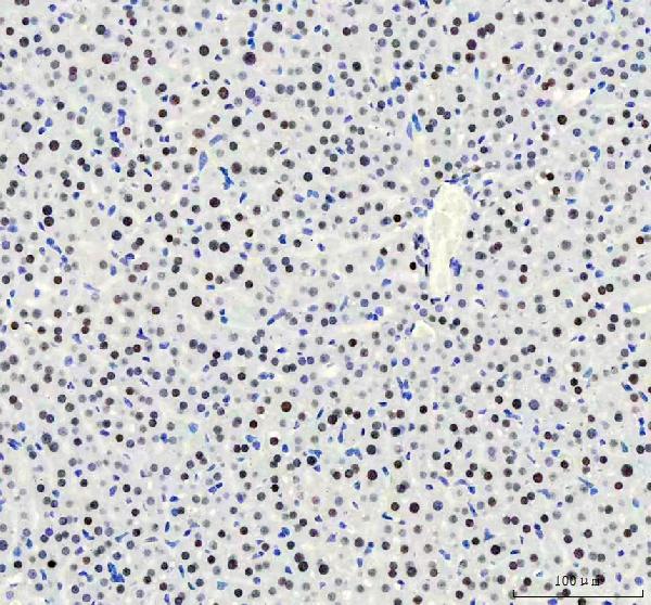
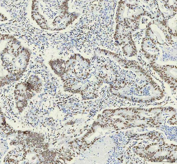
    Figure 2. IHC analysis of HNRNPM using anti-HNRNPM antibody (M06017-1).
    HNRNPM was detected in a paraffin-embedded section of human rectal cancer tissue. Heat mediated antigen retrieval was performed in EDTA buffer (pH 8.0, epitope retrieval solution). The tissue section was blocked with 10% goat serum. The tissue section was then incubated with 1:50 rabbit anti-HNRNPM Antibody (M06017-1) overnight at 4°C. Peroxidase Conjugated Goat Anti-rabbit IgG was used as secondary antibody and incubated for 30 minutes at 37°C. The tissue section was developed using HRP Conjugated Rabbit IgG Super Vision Assay Kit (Catalog # SV0002) with DAB as the chromogen.
  • 2 - Anti-HNRNPM Rabbit Monoclonal Antibody ABO16441
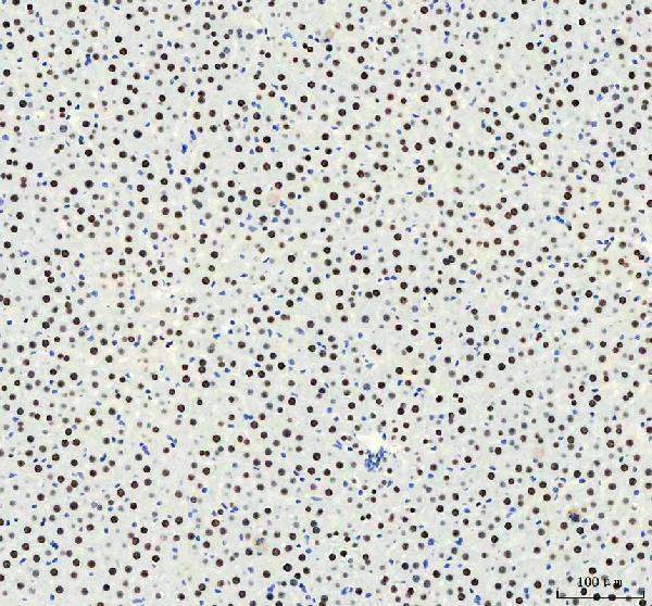
    Figure 3. IHC analysis of HNRNPM using anti-HNRNPM antibody (M06017-1).
    HNRNPM was detected in a paraffin-embedded section of human stomach cancer tissue. Heat mediated antigen retrieval was performed in EDTA buffer (pH 8.0, epitope retrieval solution). The tissue section was blocked with 10% goat serum. The tissue section was then incubated with 1:50 rabbit anti-HNRNPM Antibody (M06017-1) overnight at 4°C. Peroxidase Conjugated Goat Anti-rabbit IgG was used as secondary antibody and incubated for 30 minutes at 37°C. The tissue section was developed using HRP Conjugated Rabbit IgG Super Vision Assay Kit (Catalog # SV0002) with DAB as the chromogen.
  • 2 - Anti-HNRNPM Rabbit Monoclonal Antibody ABO16441
    Figure 4. IHC analysis of HNRNPM using anti-HNRNPM antibody (M06017-1).
    HNRNPM was detected in a paraffin-embedded section of mouse liver tissue. Heat mediated antigen retrieval was performed in EDTA buffer (pH 8.0, epitope retrieval solution). The tissue section was blocked with 10% goat serum. The tissue section was then incubated with 1:50 rabbit anti-HNRNPM Antibody (M06017-1) overnight at 4°C. Peroxidase Conjugated Goat Anti-rabbit IgG was used as secondary antibody and incubated for 30 minutes at 37°C. The tissue section was developed using HRP Conjugated Rabbit IgG Super Vision Assay Kit (Catalog # SV0002) with DAB as the chromogen.
  • 2 - Anti-HNRNPM Rabbit Monoclonal Antibody ABO16441
    Figure 5. IHC analysis of HNRNPM using anti-HNRNPM antibody (M06017-1).
    HNRNPM was detected in a paraffin-embedded section of rat liver tissue. Heat mediated antigen retrieval was performed in EDTA buffer (pH 8.0, epitope retrieval solution). The tissue section was blocked with 10% goat serum. The tissue section was then incubated with 1:50 rabbit anti-HNRNPM Antibody (M06017-1) overnight at 4°C. Peroxidase Conjugated Goat Anti-rabbit IgG was used as secondary antibody and incubated for 30 minutes at 37°C. The tissue section was developed using HRP Conjugated Rabbit IgG Super Vision Assay Kit (Catalog # SV0002) with DAB as the chromogen.
  • 产品详情
  • 实验流程
Product Information
Application
  • Applications Legend:
  • E=ELISA
  • WB=Western Blotting
  • IHC=Immunohistochemistry
  • IHC-P=Immunohistochemistry (Paraffin)
  • IP=Immunoprecipitation
  • IF=Immunofluorescence
  • IC=Immunochemistry
  • ICC=Immunocytochemistry
  • FC=Flow Cytometry
  • DB=Dot Blot
WB, IHC, IF, ICC, FC
Primary Accession P52272
Host Rabbit
Isotype IgG
Reactivity Rat, Human, Mouse
Clonality Monoclonal
Format Liquid
Description Anti-HNRNPM Rabbit Monoclonal Antibody . Tested in WB, IHC, ICC/IF, Flow Cytometry applications. This antibody reacts with Human, Mouse, Rat.
Additional Information
Gene ID 4670
Other Names Heterogeneous nuclear ribonucleoprotein M, hnRNP M, HNRNPM, HNRPM, NAGR1
Calculated MW 77516 Da
Application Details WB 1:500-1:2000
IHC 1:50-1:200
ICC/IF 1:50-1:200
FC 1:50
Source Eukaryota
Contents Rabbit IgG in phosphate buffered saline, pH 7.4, 150mM NaCl, 0.02% sodium azide and 50% glycerol, 0.4-0.5mg/ml BSA.
Clone Names Clone: 29H77
Immunogen A synthesized peptide derived from human HNRNPM
Purification Affinity-chromatography
Storage Store at -20°C for one year. For short term storage and frequent use, store at 4°C for up to one month. Avoid repeated freeze-thaw cycles.

For Research Use Only. Not For Use In Diagnostic Procedures.

Protein Information
Name HNRNPM
Synonyms HNRPM, NAGR1
Function Pre-mRNA binding protein in vivo, binds avidly to poly(G) and poly(U) RNA homopolymers in vitro. Involved in splicing. Acts as a receptor for carcinoembryonic antigen in Kupffer cells, may initiate a series of signaling events leading to tyrosine phosphorylation of proteins and induction of IL-1 alpha, IL-6, IL-10 and tumor necrosis factor alpha cytokines.
Cellular Location Nucleus, nucleolus {ECO:0000269|Ref.5}.
Research Areas
FeedBack

终于等到您。ABCEPTA(百远生物)抗体产品。
点击下方“我要评价 ”按钮提交您的反馈信息,您的反馈和评价是我们最宝贵的财富之一,
我们将在1-3个工作日内处理您的反馈信息。

如有疑问,联系:0512-88856768 tech-china@abcepta.com.


我要评价