注册 | 登录
点击这里给我发消息
所有产品
  • 所有产品
  • 一抗
  • 裂解液
>   首页   >   产品   >   一抗   >   信号转导   >   Anti-NDUFAB1 Rabbit Monoclonal Antibody   
Anti-NDUFAB1 Rabbit Monoclonal Antibody
     
  • 1 - Anti-NDUFAB1 Rabbit Monoclonal Antibody ABO16558
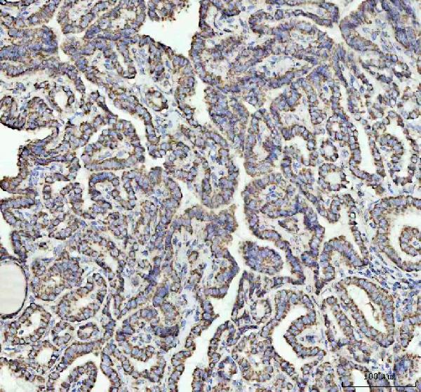
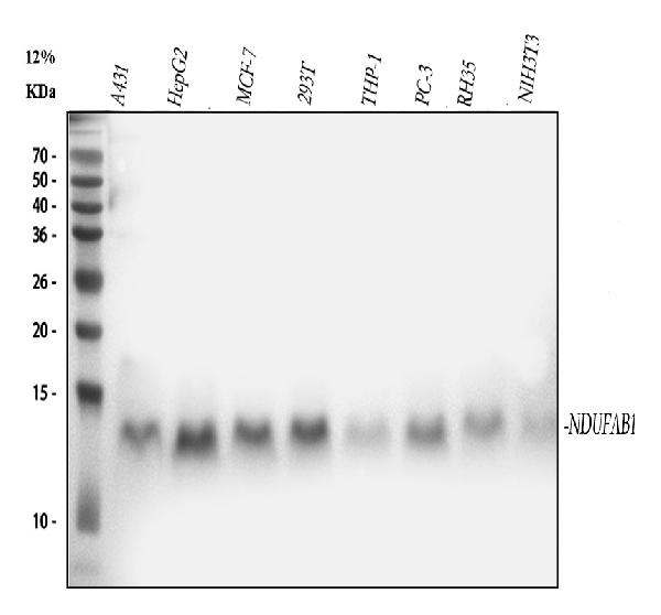
    Figure 1. Western blot analysis of NDUFAB1 using anti-NDUFAB1 antibody (M09620).
    Electrophoresis was performed on a 5-20% SDS-PAGE gel at 70V (Stacking gel) / 90V (Resolving gel) for 2-3 hours. The sample well of each lane was loaded with 30 ug of sample under reducing conditions.
    Lane 1: human A431 whole cell lysates,
    Lane 2: human HepG2 whole cell lysates,
    Lane 3: human MCF-7 whole cell lysates,
    Lane 4: human 293T whole cell lysates,
    Lane 5: human THP-1 whole cell lysates,
    Lane 6: human PC-3 whole cell lysates,
    Lane 7: rat RH35 whole cell lysates,
    Lane 8: mouse NIH/3T3 whole cell lysates.
    After electrophoresis, proteins were transferred to a nitrocellulose membrane at 150 mA for 50-90 minutes. Blocked the membrane with 5% non-fat milk/TBS for 1.5 hour at RT. The membrane was incubated with rabbit anti-NDUFAB1 antigen affinity purified monoclonal antibody (Catalog # M09620) at 1:500 overnight at 4°C, then washed with TBS-0.1%Tween 3 times with 5 minutes each and probed with a goat anti-rabbit IgG-HRP secondary antibody at a dilution of 1:500 for 1.5 hour at RT. The signal is developed using an Enhanced Chemiluminescent detection (ECL) kit (Catalog # EK1002) with Tanon 5200 system. A specific band was detected for NDUFAB1 at approximately 14 kDa. The expected band size for NDUFAB1 is at 17 kDa.
  • 2 - Anti-NDUFAB1 Rabbit Monoclonal Antibody ABO16558
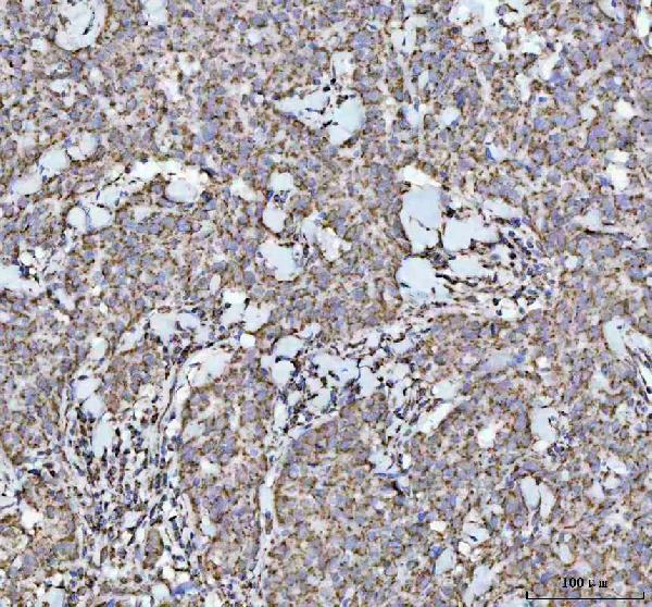
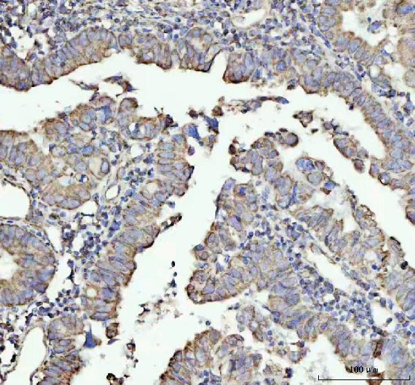
    Figure 2. IHC analysis of NDUFAB1 using anti-NDUFAB1 antibody (MM09620).
    NDUFAB1 was detected in a paraffin-embedded section of human colorectal adenocarcinoma tissue. Heat mediated antigen retrieval was performed in EDTA buffer (pH 8.0, epitope retrieval solution). The tissue section was blocked with 10% goat serum. The tissue section was then incubated with 1:50 rabbit anti-NDUFAB1 Antibody (M09620) overnight at 4°C. Peroxidase Conjugated Goat Anti-rabbit IgG was used as secondary antibody and incubated for 30 minutes at 37°C. The tissue section was developed using HRP Conjugated Rabbit IgG Super Vision Assay Kit (Catalog # SV0002) with DAB as the chromogen.
  • 2 - Anti-NDUFAB1 Rabbit Monoclonal Antibody ABO16558
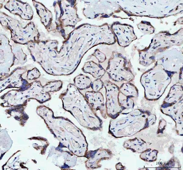
    Figure 3. IHC analysis of NDUFAB1 using anti-NDUFAB1 antibody (MM09620).
    NDUFAB1 was detected in a paraffin-embedded section of human thyroid cancer tissue. Heat mediated antigen retrieval was performed in EDTA buffer (pH 8.0, epitope retrieval solution). The tissue section was blocked with 10% goat serum. The tissue section was then incubated with 1:50 rabbit anti-NDUFAB1 Antibody (M09620) overnight at 4°C. Peroxidase Conjugated Goat Anti-rabbit IgG was used as secondary antibody and incubated for 30 minutes at 37°C. The tissue section was developed using HRP Conjugated Rabbit IgG Super Vision Assay Kit (Catalog # SV0002) with DAB as the chromogen.
  • 2 - Anti-NDUFAB1 Rabbit Monoclonal Antibody ABO16558
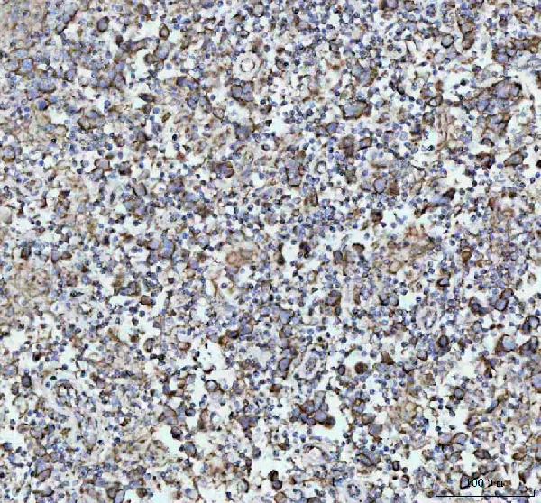
    Figure 4. IHC analysis of NDUFAB1 using anti-NDUFAB1 antibody (MM09620).
    NDUFAB1 was detected in a paraffin-embedded section of human breast cancer tissue. Heat mediated antigen retrieval was performed in EDTA buffer (pH 8.0, epitope retrieval solution). The tissue section was blocked with 10% goat serum. The tissue section was then incubated with 1:50 rabbit anti-NDUFAB1 Antibody (M09620) overnight at 4°C. Peroxidase Conjugated Goat Anti-rabbit IgG was used as secondary antibody and incubated for 30 minutes at 37°C. The tissue section was developed using HRP Conjugated Rabbit IgG Super Vision Assay Kit (Catalog # SV0002) with DAB as the chromogen.
  • 2 - Anti-NDUFAB1 Rabbit Monoclonal Antibody ABO16558
    Figure 5. IHC analysis of NDUFAB1 using anti-NDUFAB1 antibody (MM09620).
    NDUFAB1 was detected in a paraffin-embedded section of human placenta tissue. Heat mediated antigen retrieval was performed in EDTA buffer (pH 8.0, epitope retrieval solution). The tissue section was blocked with 10% goat serum. The tissue section was then incubated with 1:50 rabbit anti-NDUFAB1 Antibody (M09620) overnight at 4°C. Peroxidase Conjugated Goat Anti-rabbit IgG was used as secondary antibody and incubated for 30 minutes at 37°C. The tissue section was developed using HRP Conjugated Rabbit IgG Super Vision Assay Kit (Catalog # SV0002) with DAB as the chromogen.
  • 2 - Anti-NDUFAB1 Rabbit Monoclonal Antibody ABO16558
    Figure 6. IHC analysis of NDUFAB1 using anti-NDUFAB1 antibody (MM09620).
    NDUFAB1 was detected in a paraffin-embedded section of human testicular germ cell tumor tissue. Heat mediated antigen retrieval was performed in EDTA buffer (pH 8.0, epitope retrieval solution). The tissue section was blocked with 10% goat serum. The tissue section was then incubated with 1:50 rabbit anti-NDUFAB1 Antibody (M09620) overnight at 4°C. Peroxidase Conjugated Goat Anti-rabbit IgG was used as secondary antibody and incubated for 30 minutes at 37°C. The tissue section was developed using HRP Conjugated Rabbit IgG Super Vision Assay Kit (Catalog # SV0002) with DAB as the chromogen.
  • 产品详情
  • 实验流程
Product Information
Application
  • Applications Legend:
  • E=ELISA
  • WB=Western Blotting
  • IHC=Immunohistochemistry
  • IHC-P=Immunohistochemistry (Paraffin)
  • IP=Immunoprecipitation
  • IF=Immunofluorescence
  • IC=Immunochemistry
  • ICC=Immunocytochemistry
  • FC=Flow Cytometry
  • DB=Dot Blot
WB, IHC, IP, FC
Primary Accession O14561
Host Rabbit
Isotype IgG
Reactivity Rat, Human, Mouse
Clonality Monoclonal
Format Liquid
Description Anti-NDUFAB1 Rabbit Monoclonal Antibody . Tested in WB, IHC, IP, Flow Cytometry applications. This antibody reacts with Human, Mouse, Rat.
Additional Information
Gene ID 4706
Other Names Acyl carrier protein, mitochondrial, ACP, CI-SDAP, NADH-ubiquinone oxidoreductase 9.6 kDa subunit, NDUFAB1 (HGNC:7694)
Calculated MW 17417 Da
Application Details WB 1:500-1:2000
IHC 1:50-1:200
IP 1:50
FC 1:50
Source Eukaryota
Contents Rabbit IgG in phosphate buffered saline, pH 7.4, 150mM NaCl, 0.02% sodium azide and 50% glycerol, 0.4-0.5mg/ml BSA.
Clone Names Clone: 30N94
Immunogen A synthesized peptide derived from human NDUFAB1
Purification Affinity-chromatography
Storage Store at -20°C for one year. For short term storage and frequent use, store at 4°C for up to one month. Avoid repeated freeze-thaw cycles.

For Research Use Only. Not For Use In Diagnostic Procedures.

Protein Information
Name NDUFAB1 (HGNC:7694)
Function Carrier of the growing fatty acid chain in fatty acid biosynthesis (By similarity) (PubMed:27626371). Accessory and non- catalytic subunit of the mitochondrial membrane respiratory chain NADH dehydrogenase (Complex I), which functions in the transfer of electrons from NADH to the respiratory chain (PubMed:27626371). Accessory protein, of the core iron-sulfur cluster (ISC) assembly complex, that regulates, in association with LYRM4, the stability and the cysteine desulfurase activity of NFS1 and participates in the [2Fe-2S] clusters assembly on the scaffolding protein ISCU (PubMed:31664822). The core iron-sulfur cluster (ISC) assembly complex is involved in the de novo synthesis of a [2Fe-2S] cluster, the first step of the mitochondrial iron-sulfur protein biogenesis. This process is initiated by the cysteine desulfurase complex (NFS1:LYRM4:NDUFAB1) that produces persulfide which is delivered on the scaffold protein ISCU in a FXN- dependent manner. Then this complex is stabilized by FDX2 which provides reducing equivalents to accomplish the [2Fe-2S] cluster assembly. Finally, the [2Fe-2S] cluster is transferred from ISCU to chaperone proteins, including HSCB, HSPA9 and GLRX5 (By similarity).
Cellular Location Mitochondrion
Research Areas
FeedBack

终于等到您。ABCEPTA(百远生物)抗体产品。
点击下方“我要评价 ”按钮提交您的反馈信息,您的反馈和评价是我们最宝贵的财富之一,
我们将在1-3个工作日内处理您的反馈信息。

如有疑问,联系:0512-88856768 tech-china@abcepta.com.


我要评价