


 癌症的基本特征包括细胞增殖、血管生成、迁移、凋亡逃避机制和细胞永生等。找到癌症发生过程中这些通路的关键标记物和对应的抗体用于检测至关重要。
 为您推荐一个泛素化位点预测神器——泛素化分析工具,可以为您的蛋白的泛素化位点作出预测和评分。
 细胞自噬受体图形绘图工具为你的蛋白的细胞受体结合位点作出预测和评分,识别结合到自噬通路中的蛋白是非常重要的,便于让我们理解自噬在正常生理、病理过程中的作用,如发育、细胞分化、神经退化性疾病、压力条件下、感染和癌症。
Anti-IRBIT/AHCYL1 Antibody Picoband™ (monoclonal, 2E7D9)
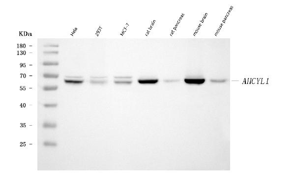
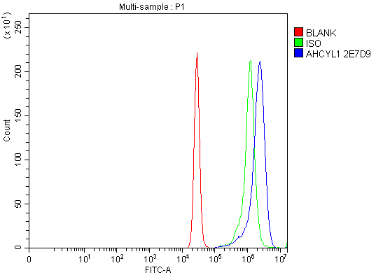
- 产品详情
- 实验流程
- 背景知识
Application 
| WB, FC |
|---|---|
| Primary Accession | O43865 |
| Host | Mouse |
| Isotype | IgG1 |
| Reactivity | Rat, Human, Mouse |
| Clonality | Monoclonal |
| Format | Lyophilized |
| Description | Anti-IRBIT/AHCYL1 Antibody Picoband™ (monoclonal, 2E7D9) . Tested in Flow Cytometry, WB applications. This antibody reacts with Human, Mouse, Rat. |
| Reconstitution | Adding 0.2 ml of distilled water will yield a concentration of 500 µg/ml. |
| Gene ID | 10768 |
|---|---|
| Other Names | S-adenosylhomocysteine hydrolase-like protein 1 {ECO:0000312|HGNC:HGNC:344}, DC-expressed AHCY-like molecule, IP(3)Rs binding protein released with IP(3), IRBIT, Putative adenosylhomocysteinase 2, S-adenosyl-L-homocysteine hydrolase 2, AdoHcyase 2, AHCYL1 (HGNC:344) |
| Calculated MW | 58951 Da |
| Application Details | Western blot, 0.25-0.5 µg/ml, Human, Mouse, Rat Flow Cytometry, 1-3 µg/1x10^6 cells, Human |
| Source | Eukaryota |
| Contents | Each vial contains 4 mg Trehalose, 0.9 mg NaCl and 0.2 mg Na2HPO4. |
| Clone Names | Clone: 2E7D9 |
| Immunogen | E.coli-derived human IRBIT/AHCYL1 recombinant protein (Position: E14-K57). |
| Purification | Immunogen affinity purified. |
| Storage | At -20°C for one year from date of receipt. After reconstitution, at 4°C for one month. It can also be aliquotted and stored frozen at -20°C for six months. Avoid repeated freezing and thawing. |
For Research Use Only. Not For Use In Diagnostic Procedures.
| Name | AHCYL1 (HGNC:344) |
|---|---|
| Function | Multifaceted cellular regulator which coordinates several essential cellular functions including regulation of epithelial HCO3(-) and fluid secretion, mRNA processing and DNA replication. Regulates ITPR1 sensitivity to inositol 1,4,5-trisphosphate, competing for the common binding site and acting as endogenous 'pseudoligand' whose inhibitory activity can be modulated by its phosphorylation status. Promotes the formation of contact points between the endoplasmic reticulum (ER) and mitochondria, facilitating transfer of Ca(2+) from the ER to mitochondria (PubMed:27995898). Under normal cellular conditions, functions cooperatively with BCL2L10 to limit ITPR1- mediated Ca(2+) release but, under apoptotic stress conditions, dephosphorylated which promotes dissociation of both AHCYL1 and BCL2L10 from mitochondria-associated endoplasmic reticulum membranes, inhibits BCL2L10 interaction with ITPR1 and leads to increased Ca(2+) transfer to mitochondria which promotes apoptosis (PubMed:27995898). In the pancreatic and salivary ducts, at resting state, attenuates inositol 1,4,5-trisphosphate-induced calcium release by interacting with ITPR1 (PubMed:16793548). When extracellular stimuli induce ITPR1 phosphorylation or inositol 1,4,5-trisphosphate production, dissociates from ITPR1 to interact with CFTR and SLC26A6, mediating their synergistic activation by calcium and cAMP that stimulates the epithelial secretion of electrolytes and fluid (By similarity). Also activates basolateral SLC4A4 isoform 1 to coordinate fluid and HCO3(-) secretion (PubMed:16769890). Inhibits the effect of STK39 on SLC4A4 and CFTR by recruiting PP1 phosphatase which activates SLC4A4, SLC26A6 and CFTR through dephosphorylation (By similarity). Mediates the induction of SLC9A3 surface expression produced by Angiotensin-2 (PubMed:20584908). Depending on the cell type, activates SLC9A3 in response to calcium or reverses SLC9A3R2-dependent calcium inhibition (PubMed:18829453). May modulate the polyadenylation state of specific mRNAs, both by controlling the subcellular location of FIP1L1 and by inhibiting PAPOLA activity, in response to a stimulus that alters its phosphorylation state (PubMed:19224921). Acts as a (dATP)-dependent inhibitor of ribonucleotide reductase large subunit RRM1, controlling the endogenous dNTP pool and ensuring normal cell cycle progression (PubMed:25237103). In vitro does not exhibit any S-adenosyl-L- homocysteine hydrolase activity (By similarity). Functions as a SAH (S- adenosyl-L-homocysteine) sensor that inhibits autophagy through an MTORC1-independent mechanism. Upon SAH binding, interacts with the catalytic domain of PIK3C3, inhibiting its lipid kinase activity and reducing production of phosphatidylinositol-3-phosphate (PtdIns3P), thereby blocking autophagosome formation (PubMed:33993848). |
| Cellular Location | Endoplasmic reticulum. Cytoplasm, cytosol. Apical cell membrane {ECO:0000250|UniProtKB:B5DFN2}; Peripheral membrane protein. Microsome {ECO:0000250|UniProtKB:Q80SW1} Note=Associates with membranes when phosphorylated, probably through interaction with ITPR1 (By similarity). Localizes to mitochondria- associated endoplasmic reticulum membranes (MAMs) (PubMed:27995898) Localization to MAMs is greatly reduced under apoptotic stress conditions (PubMed:27995898). {ECO:0000250|UniProtKB:Q80SW1, ECO:0000269|PubMed:27995898} |
| Tissue Location | Expressed in dendritic cells. |
Provided below are standard protocols that you may find useful for product applications.
BACKGROUND
Putative adenosylhomocysteinase 2 is an enzyme that in humans is encoded by the AHCYL1 gene. The protein encoded by this gene interacts with inositol 1,4,5-trisphosphate receptor, type 1 and may be involved in the conversion of S-adenosyl-L-homocysteine to L-homocysteine and adenosine. Several transcript variants encoding two different isoforms have been found for this gene.

终于等到您。ABCEPTA(百远生物)抗体产品。
点击下方“我要评价 ”按钮提交您的反馈信息,您的反馈和评价是我们最宝贵的财富之一,
我们将在1-3个工作日内处理您的反馈信息。
如有疑问,联系:0512-88856768 tech-china@abcepta.com.


