


 癌症的基本特征包括细胞增殖、血管生成、迁移、凋亡逃避机制和细胞永生等。找到癌症发生过程中这些通路的关键标记物和对应的抗体用于检测至关重要。
 为您推荐一个泛素化位点预测神器——泛素化分析工具,可以为您的蛋白的泛素化位点作出预测和评分。
 细胞自噬受体图形绘图工具为你的蛋白的细胞受体结合位点作出预测和评分,识别结合到自噬通路中的蛋白是非常重要的,便于让我们理解自噬在正常生理、病理过程中的作用,如发育、细胞分化、神经退化性疾病、压力条件下、感染和癌症。
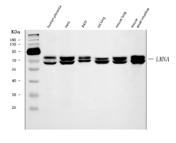
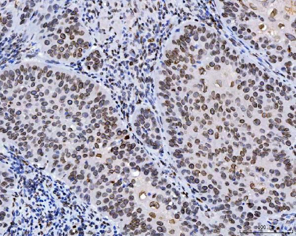
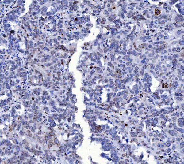
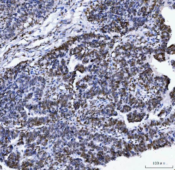
Anti-Lamin A+C/LMNA Antibody Picoband™ (monoclonal, 5F3C12)
- 产品详情
- 实验流程
- 背景知识
Application 
| WB, IHC |
|---|---|
| Primary Accession | P02545 |
| Host | Mouse |
| Isotype | IgG2b |
| Reactivity | Rat, Human, Mouse |
| Clonality | Monoclonal |
| Format | Lyophilized |
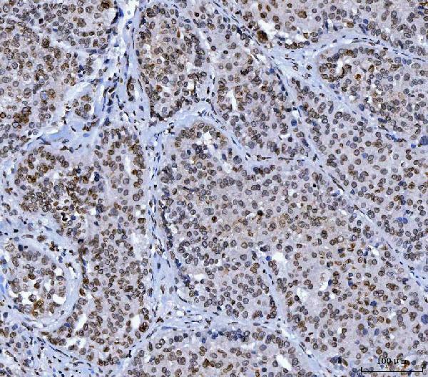
| Description | Anti-Lamin A+C/LMNA Antibody Picoband™ (monoclonal, 5F3C12) . Tested in IHC, WB applications. This antibody reacts with Human, Mouse, Rat. |
| Reconstitution | Adding 0.2 ml of distilled water will yield a concentration of 500 µg/ml. |
| Gene ID | 4000 |
|---|---|
| Other Names | Prelamin-A/C, Lamin-A/C, 70 kDa lamin, Renal carcinoma antigen NY-REN-32, LMNA, LMN1 |
| Calculated MW | 74139 Da |
| Application Details | Western blot, 0.25-0.5 µg/ml, Human, Mouse, Rat Immunohistochemistry(Paraffin-embedded Section), 2-5 µg/ml, Human |
| Source | Eukaryota |
| Contents | Each vial contains 4 mg Trehalose, 0.9 mg NaCl and 0.2 mg Na2HPO4. |
| Clone Names | Clone: 5F3C12 |
| Immunogen | E.coli-derived human Lamin A/C recombinant protein (Position: Y481-Y646). Human Lamin A/C shares 90% and 92% amino acid (aa) sequence identity with mouse and rat Lamin A/C, respectively. |
| Purification | Immunogen affinity purified. |
| Storage | At -20°C for one year from date of receipt. After reconstitution, at 4°C for one month. It can also be aliquotted and stored frozen at -20°C for six months. Avoid repeated freezing and thawing. |
For Research Use Only. Not For Use In Diagnostic Procedures.
| Name | LMNA |
|---|---|
| Synonyms | LMN1 |
| Function | [Lamin-A/C]: Lamins are intermediate filament proteins that assemble into a filamentous meshwork, and which constitute the major components of the nuclear lamina, a fibrous layer on the nucleoplasmic side of the inner nuclear membrane (PubMed:10080180, PubMed:10580070, PubMed:10587585, PubMed:10814726, PubMed:11799477, PubMed:12075506, PubMed:12927431, PubMed:15317753, PubMed:18551513, PubMed:18611980, PubMed:2188730, PubMed:22431096, PubMed:2344612, PubMed:23666920, PubMed:24741066, PubMed:31434876, PubMed:31548606, PubMed:37788673, PubMed:37832547). Lamins provide a framework for the nuclear envelope, bridging the nuclear envelope and chromatin, thereby playing an important role in nuclear assembly, chromatin organization, nuclear membrane and telomere dynamics (PubMed:10080180, PubMed:10580070, PubMed:10587585, PubMed:10814726, PubMed:11799477, PubMed:12075506, PubMed:12927431, PubMed:15317753, PubMed:18551513, PubMed:18611980, PubMed:22431096, PubMed:23666920, PubMed:24741066, PubMed:31548606, PubMed:37788673, PubMed:37832547). Lamin A and C also regulate matrix stiffness by conferring nuclear mechanical properties (PubMed:23990565, PubMed:25127216). The structural integrity of the lamina is strictly controlled by the cell cycle, as seen by the disintegration and formation of the nuclear envelope in prophase and telophase, respectively (PubMed:2188730, PubMed:2344612). Lamin A and C are present in equal amounts in the lamina of mammals (PubMed:10080180, PubMed:10580070, PubMed:10587585, PubMed:10814726, PubMed:11799477, PubMed:12075506, PubMed:12927431, PubMed:15317753, PubMed:18551513, PubMed:18611980, PubMed:22431096, PubMed:23666920, PubMed:31548606). Also involved in DNA repair: recruited by DNA repair proteins XRCC4 and IFFO1 to the DNA double-strand breaks (DSBs) to prevent chromosome translocation by immobilizing broken DNA ends (PubMed:31548606). Required for normal development of peripheral nervous system and skeletal muscle and for muscle satellite cell proliferation (PubMed:10080180, PubMed:10814726, PubMed:11799477, PubMed:18551513, PubMed:22431096). Required for osteoblastogenesis and bone formation (PubMed:12075506, PubMed:15317753, PubMed:18611980). Also prevents fat infiltration of muscle and bone marrow, helping to maintain the volume and strength of skeletal muscle and bone (PubMed:10587585). Required for cardiac homeostasis (PubMed:10580070, PubMed:12927431, PubMed:18611980, PubMed:23666920). |
| Cellular Location | Nucleus lamina. Nucleus envelope. Nucleus, nucleoplasm. Nucleus matrix. Note=Farnesylation of prelamin-A/C facilitates nuclear envelope targeting and subsequent cleavage by ZMPSTE24/FACE1 to remove the farnesyl group produces mature lamin-A/C, which can then be inserted into the nuclear lamina (PubMed:15317753) EMD is required for proper localization of non-farnesylated prelamin- A/C (PubMed:19323649). Also localizes to the micronuclear envelope in response to response to genome instability (PubMed:37788673) |
| Tissue Location | In the arteries, prelamin-A/C accumulation is not observed in young healthy vessels but is prevalent in medial vascular smooth muscle cells (VSMCs) from aged individuals and in atherosclerotic lesions, where it often colocalizes with senescent and degenerate VSMCs. Prelamin-A/C expression increases with age and disease. In normal aging, the accumulation of prelamin-A/C is caused in part by the down-regulation of ZMPSTE24/FACE1 in response to oxidative stress. |
Provided below are standard protocols that you may find useful for product applications.
BACKGROUND
Lamins are structural protein components of the nuclear lamina, a protein network underlying the inner nuclear membrane that determines nuclear shape and size. There are three types of lamins, A,B and C. The lamin A/C (LMNA) gene contains 12 exons. Alternative splicing within exon 10 gives rise to two different mRNAs that code for pre-lamin A and lamin C. Lamin A/C is mapped to 1q21.2-q21.3 and mutations in this gene cause a variety of human diseases including Emery-Dreifuss muscular dystrophy, dilated cardiomyopathy, and Hutchinson-Gilford progeria syndrome. Lamin A/C deficiency is thus associated with both defective nuclear mechanics and impaired mechanically activated gene transcription.

终于等到您。ABCEPTA(百远生物)抗体产品。
点击下方“我要评价 ”按钮提交您的反馈信息,您的反馈和评价是我们最宝贵的财富之一,
我们将在1-3个工作日内处理您的反馈信息。
如有疑问,联系:0512-88856768 tech-china@abcepta.com.


