


 癌症的基本特征包括细胞增殖、血管生成、迁移、凋亡逃避机制和细胞永生等。找到癌症发生过程中这些通路的关键标记物和对应的抗体用于检测至关重要。
 为您推荐一个泛素化位点预测神器——泛素化分析工具,可以为您的蛋白的泛素化位点作出预测和评分。
 细胞自噬受体图形绘图工具为你的蛋白的细胞受体结合位点作出预测和评分,识别结合到自噬通路中的蛋白是非常重要的,便于让我们理解自噬在正常生理、病理过程中的作用,如发育、细胞分化、神经退化性疾病、压力条件下、感染和癌症。
HRV14 3C Antibody
Rabbit Polyclonal Antibody
- 产品详情
- 实验流程
- 背景知识
Application
| WB |
|---|---|
| Primary Accession | P03303 |
| Host | Rabbit |
| Clonality | Polyclonal |
| Isotype | Rabbit IgG |
| Calculated MW | 242991 Da |
| Gene ID | 1461213 |
|---|---|
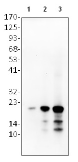
| Positive Control | WB: r HRV14 3C |
| Application & Usage | WB: 1-4 µg |
| Other Names | Precission protease, 3C Protease |
| Appearance | Colorless liquid |
| Formulation | In PBS pH 7.2, 0.01 % BSA, 0.03 % ProClin® and 50 % glycerol |
| Reconstitution & Storage | -20 °C |
| Background Descriptions | |
| Precautions | HRV14 3C Antibody is for research use only and not for use in diagnostic or therapeutic procedures. |
| Name | POLG |
|---|---|
| Function | [Capsid protein VP1]: Forms an icosahedral capsid of pseudo T=3 symmetry with capsid proteins VP2 and VP3. The capsid is 300 Angstroms in diameter, composed of 60 copies of each capsid protein and enclosing the viral positive strand RNA genome (By similarity). Capsid protein VP1 mainly forms the vertices of the capsid. Capsid protein VP1 interacts with host ICAM1 to provide virion attachment to target host cells (PubMed:10562537). This attachment induces virion internalization (By similarity). Tyrosine kinases are probably involved in the entry process. After binding to its receptor, the capsid undergoes conformational changes (By similarity). Capsid protein VP1 N-terminus (that contains an amphipathic alpha-helix) and capsid protein VP4 are externalized (Probable). Together, they shape a pore in the host membrane through which viral genome is translocated to host cell cytoplasm (PubMed:28696310). After genome has been released, the channel shrinks. |
| Cellular Location | [Capsid protein VP0]: Virion. Host cytoplasm [Capsid protein VP2]: Virion {ECO:0000250|UniProtKB:P03300}. Host cytoplasm [Capsid protein VP1]: Virion {ECO:0000250|UniProtKB:P03300}. Host cytoplasm [Protein 2C]: Host cytoplasmic vesicle membrane; Peripheral membrane protein; Cytoplasmic side. Note=Probably localizes to the surface of intracellular membrane vesicles that are induced after virus infection as the site for viral RNA replication. These vesicles are derived from the endoplasmic reticulum [Protein 3AB]: Host cytoplasmic vesicle membrane; Peripheral membrane protein; Cytoplasmic side. Note=Probably localizes to the surface of intracellular membrane vesicles that are induced after virus infection as the site for viral RNA replication. These vesicles are derived from the endoplasmic reticulum [Protease 3C]: Host cytoplasm. [RNA-directed RNA polymerase]: Host cytoplasmic vesicle membrane; Peripheral membrane protein; Cytoplasmic side. Note=Probably localizes to the surface of intracellular membrane vesicles that are induced after virus infection as the site for viral RNA replication. These vesicles are derived from the endoplasmic reticulum |
For Research Use Only. Not For Use In Diagnostic Procedures.
Provided below are standard protocols that you may find useful for product applications.
BACKGROUND
Human rhinovirus (HRV) infections are the most frequent causative agents of common cold and various other upper respiratory tract infections. Rhinoviruses are members of the picornavirus family, which have a positive-sense, single-stranded RNA genome that is translated into a single polyprotein precursor. In the case of HRVs, the viral polyprotein is mainly processed by the proteases (2A and 3C) to generate functional proteins and enzymes.
终于等到您。ABCEPTA(百远生物)抗体产品。
点击下方“我要评价 ”按钮提交您的反馈信息,您的反馈和评价是我们最宝贵的财富之一,
我们将在1-3个工作日内处理您的反馈信息。
如有疑问,联系:0512-88856768 tech-china@abcepta.com.


