


 癌症的基本特征包括细胞增殖、血管生成、迁移、凋亡逃避机制和细胞永生等。找到癌症发生过程中这些通路的关键标记物和对应的抗体用于检测至关重要。
 为您推荐一个泛素化位点预测神器——泛素化分析工具,可以为您的蛋白的泛素化位点作出预测和评分。
 细胞自噬受体图形绘图工具为你的蛋白的细胞受体结合位点作出预测和评分,识别结合到自噬通路中的蛋白是非常重要的,便于让我们理解自噬在正常生理、病理过程中的作用,如发育、细胞分化、神经退化性疾病、压力条件下、感染和癌症。
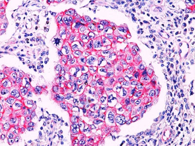
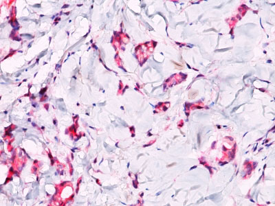
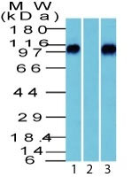
beta-Catenin (p120) Antibody - With BSA and Azide
Rabbit Polyclonal Antibody
- 产品详情
- 实验流程
- 背景知识
Application
| WB, IF, FC, IHC-P |
|---|---|
| Primary Accession | P35222 |
| Other Accession | 1499, 476018 |
| Reactivity | Human, Mouse |
| Host | Rabbit |
| Clonality | Polyclonal |
| Isotype | Rabbit / IgG |
| Clone Names | |
| Calculated MW | 92 KDa |
| Other Names | Catenin beta-1, Beta-catenin, CTNNB1, CTNNB |
|---|---|
| Application Note | WB~~1:1000 IF~~1:50~200 FC~~1:10~50 IHC-P~~1:50~200 |
| Format | 200ug/ml of Ab purified from rabbit anti-serum by Protein A. Prepared in 10mM PBS with 0.05% BSA & 0.05% azide. Also available WITHOUT BSA at 1.0mg/ml. |
| Storage | Store at 2 to 8°C.Antibody is stable for 24 months. |
| Precautions | beta-Catenin (p120) Antibody - With BSA and Azide is for research use only and not for use in diagnostic or therapeutic procedures. |
For Research Use Only. Not For Use In Diagnostic Procedures.
Provided below are standard protocols that you may find useful for product applications.
BACKGROUND
Beta-catenin associates with the cytoplasmic portion of E-cadherin, which is necessary for the function of E-cadherin as an adhesion molecule. In normal tissues, beta-catenin is localized to the membrane of epithelial cells, consistent with its role in the cell adhesion complex. In breast ductal neoplasia, beta-catenin is usually localized in cellular membranes. However, in lobular neoplasia, a marked redistribution of beta-catenin throughout the cytoplasm results in a diffuse cytoplasmic pattern. Immuno-staining of beta-catenin and E-cadherin is helps in the accurate identification of ductal and lobular neoplasms, including a distinction between low-grade ductal carcinoma in situ (DCIS) and lobular carcinoma. Additionally, some rectal and gastric adenocarcinomas demonstrate diffuse cytoplasmic beta-catenin staining and a lack of membranous staining, mimicking the staining pattern observed with lobular breast carcinomas.
REFERENCES
Dabbs DJ et. al. Am J Surg Path. 2007;31:427-437. | Sarrio D et. al. Oncogene. 2004;23:3272-3283. | Mastracci TL et. al. Mod Path. 2005;18:741-751
终于等到您。ABCEPTA(百远生物)抗体产品。
点击下方“我要评价 ”按钮提交您的反馈信息,您的反馈和评价是我们最宝贵的财富之一,
我们将在1-3个工作日内处理您的反馈信息。
如有疑问,联系:0512-88856768 tech-china@abcepta.com.


