


 癌症的基本特征包括细胞增殖、血管生成、迁移、凋亡逃避机制和细胞永生等。找到癌症发生过程中这些通路的关键标记物和对应的抗体用于检测至关重要。
 为您推荐一个泛素化位点预测神器——泛素化分析工具,可以为您的蛋白的泛素化位点作出预测和评分。
 细胞自噬受体图形绘图工具为你的蛋白的细胞受体结合位点作出预测和评分,识别结合到自噬通路中的蛋白是非常重要的,便于让我们理解自噬在正常生理、病理过程中的作用,如发育、细胞分化、神经退化性疾病、压力条件下、感染和癌症。
GFAP (Astrocyte & Neural Stem Cell Marker) Antibody - With BSA and Azide
Mouse Monoclonal Antibody [Clone SPM507 ]
- 产品详情
- 实验流程
- 背景知识
Application
| WB, IF, FC, IHC-P |
|---|---|
| Primary Accession | P14136 |
| Other Accession | 2670, 514227 |
| Reactivity | Human, Mouse, Rat, Rabbit, Pig, Chicken, Bovine |
| Host | Mouse |
| Clonality | Monoclonal |
| Isotype | Mouse / IgG1 |
| Clone Names | SPM507 |
| Calculated MW | 49880 Da |
| Gene ID | 2670 |
|---|---|
| Other Names | Glial fibrillary acidic protein, GFAP, GFAP |
| Application Note | WB~~1:1000 IF~~1:50~200 FC~~1:10~50 IHC-P~~1:50~200 |
| Format | 200ug/ml of Ab purified from Bioreactor Concentrate by Protein A/G. Prepared in 10mM PBS with 0.05% BSA & 0.05% azide. Also available WITHOUT BSA & azide at 1.0mg/ml. |
| Storage | Store at 2 to 8°C.Antibody is stable for 24 months. |
| Precautions | GFAP (Astrocyte & Neural Stem Cell Marker) Antibody - With BSA and Azide is for research use only and not for use in diagnostic or therapeutic procedures. |
| Name | GFAP |
|---|---|
| Function | GFAP, a class-III intermediate filament, is a cell-specific marker that, during the development of the central nervous system, distinguishes astrocytes from other glial cells. |
| Cellular Location | Cytoplasm. Note=Associated with intermediate filaments |
| Tissue Location | Expressed in cells lacking fibronectin. |
For Research Use Only. Not For Use In Diagnostic Procedures.
Provided below are standard protocols that you may find useful for product applications.
BACKGROUND
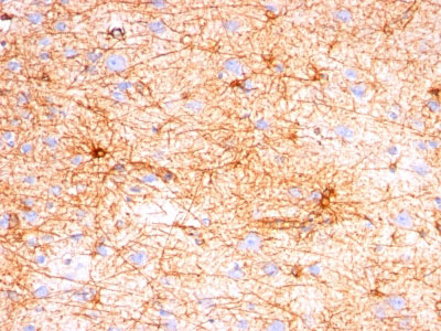
This MAb recognizes a protein of ~50kDa which is identified as Glial Fibrillary Acidic Protein (GFAP). It shows no cross-reaction with other intermediate filament proteins. GFAP is specifically found in astroglia. GFAP is a very popular marker for localizing benign astrocyte and neoplastic cells of glial origin in the central nervous system. Antibody to GFAP is useful in differentiating primary gliomas from metastatic lesions in the brain and for documenting astrocytic differentiation in tumors outside the CNS.
REFERENCES
Herpers MJ et. Al. 1986, Acta Neuropathol, 70:333-339. | Van Muijen GN et. al. 1987, Lab Invest, 57:359-369. | Debus E, et. al. 1983, Differentiation, 25(2):193-203
终于等到您。ABCEPTA(百远生物)抗体产品。
点击下方“我要评价 ”按钮提交您的反馈信息,您的反馈和评价是我们最宝贵的财富之一,
我们将在1-3个工作日内处理您的反馈信息。
如有疑问,联系:0512-88856768 tech-china@abcepta.com.


